A new label-free microscopic approach that allows scientists to observe the formation and evolution of cell membrane focal adhesions has been developed. In a paper published earlier this week in Light: Science & Applications, researchers at the Beckman Institute for Advanced Science and Technology and the Micro and Nanotechnology Laboratory at the University of Illinois explain their novel live cell imaging technique called Photonic Resonator Outcoupler Microscopy (PROM).
"This is a new kind of biophysics method used to measure the peak intensity shift (PIS) of the spectra reflected from the biomaterials on a photonic crystal surface," said Yue Zhuo, a Beckman Institute postdoctoral fellow and first author on the paper. "The PIS indicates the variation of cluster size in the focal adhesion area of the cell while it's alive."
"Typically people look at focal adhesions with fluorescent tags or proteins," Zhuo said. "But fluorescent imaging is an invasive imaging method that may change the conformations or block the binding sites of the proteins in the focal adhesion area." Fluorescence microscopy is also severely limited by photobleaching, in which the fluorophores only maintain their brightness for several seconds. Because PROM does not use fluorophores, and only uses low-intensity illumination, there is not a limit to how long live cells can be measured.
PROM utilizes a photonic crystal biosensor surface to create an evanescent field, which selectively illuminates only the ECM-attached cell membrane and associated protein aggregates directly inside the cell membrane. The photonic crystal strictly limits lateral propagation of light while keeping light tightly bound to the biosensor surface, to enable high-resolution imaging of the cell membrane attachment footprint.
"PROM is providing real-time information about dynamic processes that occur specifically on cell membranes that is not available by any other method," said Brian Cunningham, a professor of electrical and computer engineering and bioengineering, and the principle investigator for the PROM project. "Since so many biological processes are mediated through attachment of cells to surfaces, PROM provides a unique view of migration, chemotaxis, chemotoxicity, differentiation, biofilm formation, and division. We see PROM as an exciting new tool for cell biologists that can also be applied towards personalized anticancer drug selection, tissue engineering, and sensor-integrated tumor modeling."
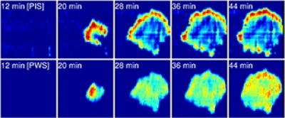
Image: A photonic Resonator Outcoupler Microscopy (PROM) image that highlights focal adhesions of live dental stem cells. Image courtesy of Yue Zhuo/Brian Cunningham.