Thanks to the cerebrospinal fluid, the thought of being hungry comes to mind. Work from researchers at the University of Southern California has shown that the cerebrospinal fluid helps send molecules to the brain to give you an appetite. The work was published yesterday in Cell Metabolism.
"People usually think of brain cells as communicating signals through the synapses between them," said first author Emily Noble. "We are showing that the brain has another complementary way to communicate by sending these signals into the cerebrospinal fluid."
"The cerebrospinal fluid had been historically thought more of as a metabolic wasteland," said Scott Kanoski, the study's corresponding author. "But what we are showing is that the fluid is an active mechanism for communication in the brain."
In this study, the team focused on the melanin-concentrating hormone (MCH) molecule, which is responsible for stimulating appetite and slowing energy expenditure. Through a number of experiments, the researchers stimulated the release of the hunger peptide and tracked it in the cerebrospinal fluid.
"When we released MCH into the cerebrospinal fluid, the animals would start eating," Kanoski said. "When we reduced the levels of the molecule, then we saw the opposite effect and the animals would eat less."
From their findings, the researchers have concluded that the peptide's release is influenced by a circadian clock and mealtime routine.
The team now hopes to look into the following questions for their next steps:
Is MCH released from the brain in some special form that protects it from damage or other degradation?
How exactly does it travel into the cerebrospinal fluid and where does it go from there?
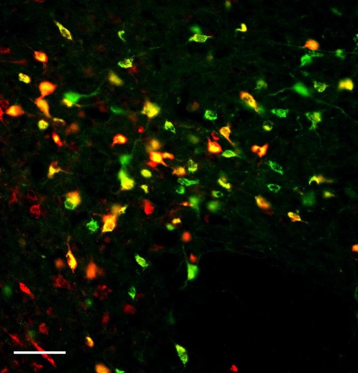 Image: Cells that communicate with the cerebrospinal fluid are marked with a red fluorescent tracer, whereas cells that make the appetite-promoting neuropeptide melanin-concentrating hormone (MCH) are shown with a green fluorescent marker. This shows that many MCH cells communicate with cerebrospinal fluid (yellow cells), the first clue that MCH may increase feeding through cerebrospinal fluid signaling. Image courtesy of Kanoski lab at the USC Dornsife College.