A new study using self-assembling peptides demonstrates that beta-peptides can be attached to an organic molecule to form a robust biomaterial that could be used in variety of biomedical applications. The researchers used a beta-peptide based structure as a bioscaffolding to successfully grow functionally active neurons. The study was published in APL Bioengineering.
Beta-peptides are composed entirely of beta amino acids which have a second carbon atom, unlike their alpha counterpart, which enables them to be resistant against proteolytic degradation. Beta-peptides can self-assemble if the amino end of the peptide is capped and can be combined with other molecules to create beta-foldamers. The study shows for the first time that beta-peptides could be attached to organic molecules that have previously been a challenge for researchers to work with.
"We were quite surprised that a very small peptide was able to still assemble, despite the fact that there was something in the middle of it," said Mark Del Borgo, an author on the paper.
"One cool thing about these is that they are completely sequence-independent," Del Borgo said. "No matter how they are made up, they assemble entirely on their own."
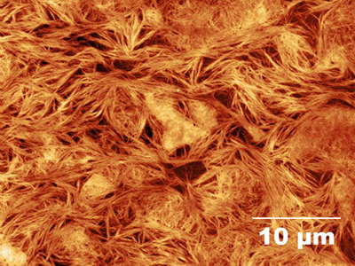
The research team created a beta-foldamer with arginylglycylaspartic acid (RGD), an alpha-peptide, as the linking molecule at the center of the complex. RGD is found in the extracellular matrix and has fibronectin-like activity. A mesh was constructed from the RGD-based beta-foldamer on which the bioengineers were able to culture a network of neurons. Not only did the neurons grow, they were able to communicate with each other as they would in a normal human brain.
 A bioscaffold on which brain cells can grow could be a beneficial for treating stroke or traumatic brain injury sufferers. Next, the research team plans to look at how these bioscaffolds could be used in a mouse model of brain injury.
Image: Beta peptide-based structures might provide the bioscaffolding for brain meshes that can help coordinate the growth of neurons after a patient experiences a stroke or traumatic brain injury. This image shows a topographical view of the mesh capable of growing neurons. Image courtesy of Mark Del Borgo.