Nicotine is a human teratogen linked to prenatal morbidity, sudden infant death, attention deficit hypersensitivity disorder (ADHD), among other deficits in cognitive function, learning, and memory. Past studies have suggested that these defects result from nicotine-induced depolarization of key ion channels and subsequent disruption of bioelectrical signaling in the embryonic cells.
A new study now demonstrates that such signals can be predicted, mapped, and manipulated to rescue developmental brain damage. The published findings in Nature Communications come from a research team at Tufts University.
 Study senior author Michael Levin provides perspective, saying "studies focusing on gene expression, growth factors, and molecular pathways have provided us with a better but still incomplete understanding of how cells arrange themselves into complex organ systems in a growing embryo."
To study bioelectrical signaling, Levin and his team used an open-source 2D computational simulation platform, called the BioElectric Tissue Simulation Engine (BETSE) to create a dynamic map of voltage signatures in a developing frog embryo. Drawing from molecular data on ion channel behaviors, the BETSE model can accurately replicate the distinct pattern of membrane voltage observed in normal embryonic brain development. More importantly, voltage mapping by BETSE could measure the "flattened," or erased, electrical pattern that is observed from nicotine exposure.
Using the BETSE model, the team tested the effects of various compounds on the embryonic voltage map. In particular, adding the protein HCN2 (hyperpolarization-activated cyclic nucleotide gated channel) to cells selectively showed enhanced hyperpolarization in areas where it was otherwise diminished by nicotine.
In vivo experiments in live embryos then further confirmed that expression of HCN2 effectively rescues the effects of nicotine exposure. The team observed restoration of the normal bioelectric pattern, brain morphology, gene expression markers, and near-normal learning capacity in the grown tadpole.
"We are now beginning to see how electrical patterns in the embryo are guiding large scale patterns of tissues, organs, and limbs. If we can decode this electrical communication between cells, then we might be able to use it to normalize development or support regeneration in the treatment of disease or injury," says Levin.
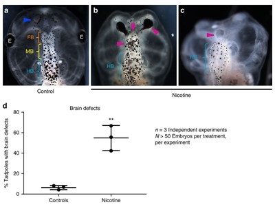
Image: Figure demonstrating how nicotine induces brain morphology defects in Xenopus embryos. Image courtesy of Michael Levin and Springer Nature.