A novel way to rapidly detect metabolically active Mycobacterium tuberculosis in resource-limited environments has been developed. Using a color-changing dye based on trehalose, a sugar that makes up outer membrane of M. tuberculosis, scientists from the University of the Witwatersrand say TB patients can now be more accurately and quickly diagnosed and treated.
The innovation involved the fusing of trehalose with a DMN molecule. The resultant DMN-trehalose is then fed to sputum-derived bacteria. The TB bacteria take it up and incorporate the DMN-trehalose into their own cell walls. The bacteria then light up under a microscope when illuminated with fluorescent light.
DMN-trehalose is naturally fluorescent and color changing, and it only illuminates in the presence of specifically TB-type bacteria. The advantage is there is no background signal indicating anything other than TB-type bacteria and thus no need to wash to reveal these bacteria, according to the team. Additional details on the new process can be found in a paper published yesterday in Science Translational Medicine.
"Where we're positioning the new stain is to create a simpler process, which provides the opportunity to stain TB bacteria in a smear more quickly and with high specificity," says Bavesh Kana, head of the Centre of Excellence in Biomedical TB Research at Wits and co-author of the paper.
TB bacteria have traditionally been detected in three ways: sputum smear staining microscopy; culturing tubercle bacteria in a laboratory, which takes up to 42 days; and detection of the DNA of TB bacteria using the GeneXpert machine. The new DMN-trehalose innovation aims to make smear microscopy easier to perform.
The new DMN-trehalose stain also enables monitoring of a patient's response to treatment.
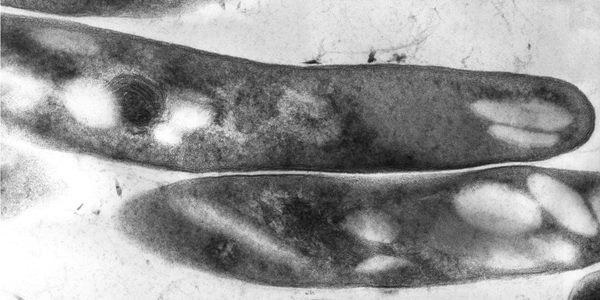
Image: Mycobacterium tuberculosis is the bacteria that cause tuberculosis (TB). Image courtesy of University of the Witwatersrand.