A research team has created a novel cell-based array for screening interactions between glycans and proteins. This new technique avoids the high cost and long turnaround times associated with generating traditional glycan microarrays. The study findings were published in Nature Communications.
Glycans are known to possess diverse biological function. These saccharide molecules of various lengths are linear or branched in structure and mostly reside on cell membranes empowering them to have important roles in signal transduction and cell recognition. They mediate interactions between two different cells and between cells and other molecules, usually through interaction with a receptor, lectin, or enzyme.
Traditionally, to study which proteins in a cell interact with glycans, scientists hybridize their cell or protein with an array of a number of glycans and observe which proteins bind to which glycans. However, this process is technically challenging, time-consuming, and expensive.
"In the past, if you wanted to make an array with 100 sugars, then you had to chemically synthesize 100 sugars individually, which can be difficult," says Peng Wu, PhD, a TSRI associate professor and senior author of the study. "Only specialized carbohydrate chemists can make them in certain labs."
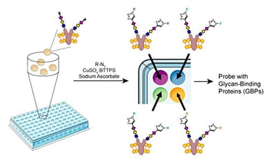 The research team started with an engineered Chinese hamster ovary (CHO) cell line, Lec2, which naturally had simple structured glycans on the cell surface. The cells were mixed with a variety of enzymes that work together to add carbohydrate branches and even unnatural sugars to each glycan. The result was an in-solution cell-based glycan array with a diverse number of glycan molecules. The total time to create the library was only 2 to 3 hours as opposed to days using traditional methods.
The cell-based array was tested and three glycan molecules bound strongly to Siglec-15, a known glycan-binding protein that is a potential drug target for osteoporosis therapies. To further validate their findings, the team mixed human osteoprogenitor cells with CHO cells displaying one of the three identified glycans and showed that the formation of Siglet-15 expressing osteoclasts was suppressed. This test provides a good indication that Siglet-15 is a promising osteoporosis target and that the glycan screen could help researchers identify potential new drug targets.
Image: A new method for assembling a glycan array, courtesy of the Wu Lab / The Scripps Research Institute.