The release of mitochondrial DNA (mtDNA) out into surrounding cellular regions is known to trigger inflammatory and immune responses. mtDNA signaling has since been implicated in autoimmune diseases such as lupus. However, the mechanisms of mtDNA release have remained unclear. Findings now reveal for the first time, through advanced microscopy techniques, the progression of mtDNA release in real-time.
 "This has been a brilliant collaboration—between Monash's Biomedicine Discovery Institute, the Walter and Eliza Hall Institute of Medical Research here in Melbourne and the Janelia Research Campus in the US—which has brought together cutting-edge technologies and first-class expertise to address questions that before now, had never been asked, and would have been impossible to answer," said senior author Benjamin Kile.
According to their published paper in Science, the team used a combination of live-cell lattice light-sheet microscopy (LLSM), 3D structured illumination microscopy, correlative light electron microscopy, and electron cryotomography to investigate mtDNA release within the intrinsic apoptosis pathway, the classical form of programmed cell death.
They found that once the pro-apoptotic proteins BAK and BAX were activated, the mitochondrial network began to break down. BAK and BAX then appeared in the mitochondrial outer membrane to form pores. These pores ultimately caused the inner membranes to lose integrity and allowed the mtDNA to escape into the cytoplasm.
"What we witnessed in real time was these professional killer proteins opening up huge 'macropores' in the outer membrane of the mitochondria, leading the inner contents to herniate out, bringing the mtDNA with it," Kile elaborated. "BAK and BAX deliver the 'kill shot' designed to permanently disable the cell. But in doing that, mtDNA is lost from the mitochondria.”
“In essence, this is collateral damage, which, if it isn't controlled properly, triggers the immune system to drive pathological inflammation, Kile adds. “Fundamental discoveries such as this are rare, and this one has profound implications for the understanding of a wide range of autoimmune diseases and infections."
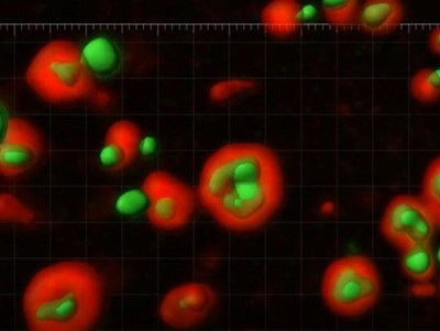
Image: A video depicts mtDNA (green) escaping the mitochondria (red) during cell death. Image courtesy of Kate McArthur, Lachlan Whitehead and the Advanced Imaging Centre at Janelia Research Campus.