A novel bioprinting technique that combines molecular self-assembly and additive manufacturing to generate constructs with tunable molecular composition and structural control has been developed. According to the Queen Mary University scientists behind this development, this technique has broad utility in areas like tissue engineering, in vitro models, and drug screening.
The constructs, made using cells and molecules normally found in natural tissues, are embedded in an “ink” that is similar to their native environment. As a result, researchers can observe how cells work within these environments and learn more about where cancer grows or how immune cells interact with other cells, for example.
The technique, which was published today in Advanced Functional Materials, integrates the micro- and macroscopic control of structural features that printing provides with the molecular and nano-scale control enabled by self-assembly. Because of this, it addresses a major need in 3D printing where commonly used printing inks have limited capacity to actively stimulate the cells that are being printed.
“This method enables the possibility to build 3D structures by printing multiple types of biomolecules capable of assembling into well-defined structures at multiple scales,” explains PhD student Clara Hedegaard, leading author of the paper. “Because of this, the self-assembling ink provides an opportunity to control the chemical and physical properties during and after printing, which can be tuned to stimulate cell behavour."
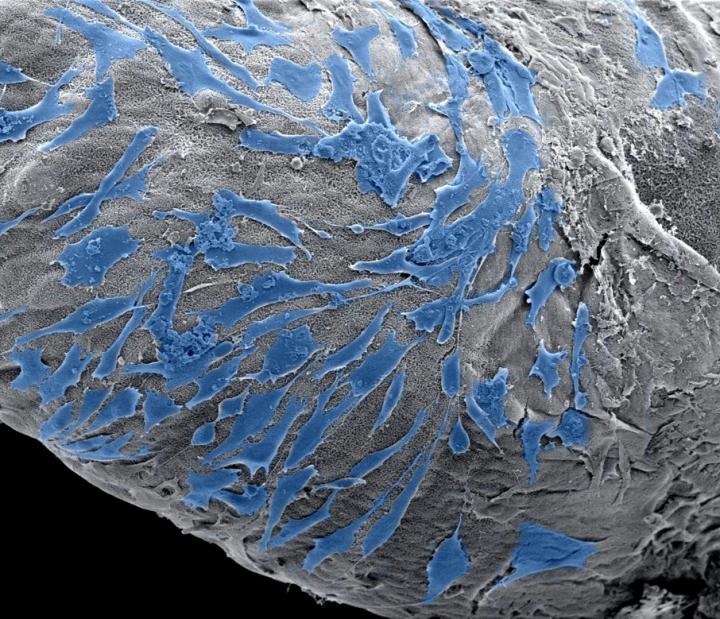
Image: Cells spreading on the outside of a peptide amphiphiles based scaffold. Image courtesy of Clara Hedegaard.