Researchers from the Francis Crick Institute have introduced a new method for near-complete labeling of all cells within a brain structure. The team reports today in Nature Communications the use of nanoengineered microelectrodes to reveal the architecture of the mouse olfactory glomerulus.
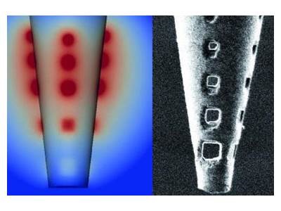 "Traditionally, scientists have either used colour-tagged viruses or charged dyes with an applied electric current to stain brain cells, but these approaches either don't label all cells or they damage the surrounding tissue," said principal study author Andreas Schaefer.
In their paper, the team described “the development of nanoengineered electroporation microelectrodes (NEMs).” A key feature of the method involved creating a series of tiny microscopic holes near the bottom tip of a glass micropipette. Through this, the team found that they could still use charged dyes but distribute the electrical current over a wider area to stain cells without damaging them.
Unlike methods that use viral vectors, application of the dyes through the NEM microcircuit stained up to 100% of all cells. And compared to conventional glass capillaries, use of a micropipette also allows for larger volumes that are configurable to the geometry of the target circuit.
The team’s paper concludes that “by relying solely on targeting an electrode to the region of interest and passive biophysical properties largely common across cell types, this can easily be employed anywhere in the central nervous system.”
"We're obviously working at a really small scale, but as the brain is made up of repeating units, we can learn a lot about how the brain works as a computational machine by studying it at this level,” says Schaefer. “Now that we have a tool of mapping these tiny units, we can start to interfere with specific cell types to see how they directly control behaviour and sensory processing."
Image: Model (left) and high resolution image (right) of the nanoengineered micropipette with holes to distribute electrical current. Image courtesy of Daniel Schwarz.