Within the brain, proteins known as ionotropic glutamate receptors (iGluRs) mediate neurotransmission at the majority of neuronal synaptic connections. Binding of glutamate activates iGluR and triggers excitatory synaptic signals. However, previous structural and computational data on iGluRs do not yet comprehensively explain how glutamate reaches the binding site of the receptor. Now, a Johns Hopkins-led team of researchers have charted an atomic scale map that tracks glutamate’s physical pathway to this receptor.
"All of this happens within a millisecond, and what hasn't been known is the way receptors latch onto glutamate,” says senior investigator Albert Lau. “Our new experiments suggest that glutamate molecules need to take very particular pathways on the surface of glutamate receptors in order to fit into a pocket within the receptor."
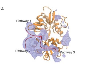 The team carried out an experiment to analyze the dynamics of a truncated version of iGluR and glutamate in solution. Interactions of nearly 50,000 atoms were then recorded and analyzed using Anton, a supercomputer run by the Pittsburgh Supercomputing Center. By counting how frequently glutamate fits in every position on the receptor, the team discovers that glutamate spends most of its time gliding into three distinct pathways.
A closer look at the pathways reveal that the glutamate’s negatively charged atoms are guided by positively charged atoms on the neuron's glutamate receptors. "What we see is an electrostatic connection, and the path glutamate follows is determined by where the charges are," says Lau.
According to the team’s paper published in Neuron, the charged residues allow glutamate to traverse a more simplified two-dimensional path to the binding pocket, rather than one in three-dimensions. Furthermore, glutamate also appears to bind in an inverted conformation, as well as reorient while in its pocket.
In cellular electrophysiological experiments with collaborators from Humboldt University, the team mutated the glutamate receptor to revert positively charged residues into either negatively charged or uncharged ones. In measuring the resulting electrical currents, the team found that mutated glutamate receptors activated at a slower rate—only at half the speed.
"If, as we think is the case, communication between neurons has to happen at a particular rate for effective brain activity, then slowing down that rate means that the brain won't work as well," says Lau. "We believe that these glutamate receptors have evolved a way to speed up the binding process."
The team’s paper concludes that the principles of electrostatic interactions as a means of funneling ligands may be a feature shared by other neurotransmitter receptors.
Image: Generated model of glutamate binding pathways. Image courtesy of Albert Y. Lau / Neuron / Elsevier.