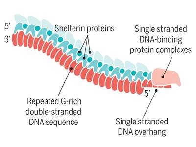
A protein complex known as “shelterin” functions as a protective shield for vertebrate telomeres, ensuring that a cell can continue to divide. For that reason, the role of shelterins have been considered in therapeutic strategies for cancer. Recent findings have now further elucidated the signaling system that drives regulation of TRF1, an essential component of the shelterin complex. Researchers from the Telomeres and Telomerase Group at the Spanish National Cancer Research Centre have led this study.
"The idea was to look for drugs that were able to reduce levels of TRF1, one of the essential shelterin proteins for the integrity of telomeres", said senior author Maria A. Blasco. "We found several that, when administered, caused damage in these structures and this led to the cancer cells not being able to divide, but we did not know what their precise target was".
 This reduction of TRF1, the team finds, is due to the phosphatidylinositol-3-kinase (PI3K) pathway. PI3K is a family of enzyme notable for its role in controlling cell proliferation, survival, and cell growth. Its downregulation has also been shown to extend life-span in organisms from yeast to mice. According to team’s report published in Nature Communications, inhibition of PI3K using specific inhibitors also inhibits its downstream target, the kinase AKT, which phosphorylates TRF1.
In a Western Blot experiment, the team shows that PI3K inhibitors cause a loss of AKT phosphorylation at select sites. This results in inhibition of TRF1 foci, visible by immunofluorescence of treated cell lines. Western analyses likewise show decreased protein expression of TRF1 following inhibitor treatment.
"We then studied whether AKT modified TRF1 in any way and we saw, through different experiments, that this was indeed the case,” reported Blasco. “AKT also modified TRF1 by phosphorylation". In vitro phosphorylation assays of TRF1 followed by tandem mass spectrometry (MS/MS) have shown that TRF1 is directly activated by AKT at select amino acid residues. Engineered variants of TRF1 lacking these key sites further confirm abnormal TRF1 phosphorylation and loss of TRF1 foci in cells. Finally, experiments in a mouse models for PI3Kα-specific inhibition confirm an in vivo decrease in TRF1 along with increased DNA damage.
Essentially, by blocking PI3K, these TRF1 is not phosphorylated and becomes unstabilized. It becomes less bound to telomeres, thus resulting in more DNA damage. The team’s findings relate the functions of PI3K and AKT with telomere stability, effectively connecting two of the major pathways for cancer and aging. In addition to P13K-class antitumour drugs, targeting TRF1 directly may provide new avenue for therapy.
Image: Simplified schematic of the human telomere complex consisting of a chromosomal-terminal segment of a tandemly repeated DNA sequence bound by protective shelterin component proteins. Image courtesy of Elizabeth H. Blackburn / Science.