UCLA researchers have described methods to produce an optimized brain organoid in vitro. This “mini brain” reproducibly produces brain structures similar to that of a living human brain. Using this model, the team has been able to study Zika infection and identify targets for treatment.
Over the past few years, scientists have faced a number of challenges in producing suitable stem cell-derived organoids that can effectively mimic the human brain. These issues include inconsistent cellular composition, failure to mimic the brain’s layered structure, inadequate size, and short lifespan.
With some key modifications, the UCLA team has been able to overcome some of these hurdles. They have used a specific number of stem cells and specialized petri dishes with a modified chemical environment. They have also added a growth factor called LIF, which stimulates a cell-signaling pathway that is critical for human brain growth. Their optimized organoids now grow larger, survive longer, and feature a stratified structure similar to the brain’s onion-like layers.
"While our organoids are in no way close to being fully functional human brains, they mimic the human brain structure much more consistently than other models," said Momoko Watanabe, a UCLA postdoctoral fellow and the study's first author. "Other scientists can use our methods to improve brain research because the data will be more accurate and consistent from experiment to experiment and more comparable to the real human brain."
The team then exposes these organoids to Zika and discover specifically how the virus destroys neural stem cells, the brain’s precursor in fetal development. By immunostaining for the Zika envelope protein, they have found four organoid surface receptors that make it possible for the virus to infect the cells. Staining with cell viability dyes then shows widespread apoptotic death for neural progenitor cells.
In addition, RNA sequencing of transcripts show suppression of genes associated with neural development, differentiation, and morphogenesis. Mapping these changes in the neural stem cells present a clearer picture of how the virus infiltrates and harms fetal brain tissue.
Testing several drugs on the Zika-infected organoids, the team identify three that are particularly effective in blocking viral entry into the brain tissue. Two of these protect neural stem cells by preventing the interaction between the virus and the entry receptors. One of these drugs have already shown promise in a previous study in reducing Zika-induced brain damage in fetal mice.
In addition to Zika, the research team plans to continue using this organoid model to better understand human brain development and neurological conditions such as autism, epilepsy, and neurodegeneration. "Many neurological diseases or conditions arise from defects in the way one neuron communicates with another or from the way an external factor, such as a virus, interacts with neural cells," says Bennett G. Novitch, the principal investigator. "If we can focus in at the level of cellular communication, we should be able to model those undesirable cellular interactions and counteract them with drugs or other therapies."
The study was published yesterday in Cell Reports.
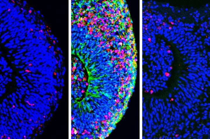
Image: Organoids before (left) and after exposure to Zika (center), and after treatment (right). Image courtesy of UCLA Broad Stem Cell Research Center/Cell Reports.