Sequencing of DNA or RNA molecules is often done by instruments that require high quantities of nucleic acid fragments. Except for third-generation sequencing platforms such as Oxford Nanopore and PacBio, a DNA amplification step is included in and/or before the sequencing run.
Interestingly, each sequencing technology and era has its own amplification and signal-generation strategy.
Next-generation sequencers Search Now Search our directory to find the right next-generation sequencers for your research needs.
This article will cover the main forms of DNA amplification and how they are coupled with different sequencing platforms, from PCR for early Sanger sequencing, to developments in fourth-generation sequencing that preserve spatial resolution.
First-generation sequencing: PCR and Sanger sequencing
PCR has become the DNA amplification method used with first-generation sequencing. At first, these methods relied on cloned DNA fragments, but PCR made the workflow easier and allowed the sequencing of many other low-quantity samples. The PCR process is considered to be straightforward at least compared to newer methods.
- DNA, primers against a target amplicon, dNTPs, and heat-resistant TaqPol are added together.
- A thermocycler makes samples go through denaturation, annealing, and extension cycles, exponentially increasing the quantity of target DNA.
This enriches a short sequence of DNA that can now be sequenced. It is used for low-DNA samples before Sanger sequencing, which uses chain-termination for detection. In Sanger:
- The DNA to be sequenced is mixed with one primer, DNA polymerase, normal dNTPs, and fluorescent ddNTPs.
- Then, polymerase extends the strand until a ddNTP is incorporated, which stops synthesis.
- This creates a mixture of fragments of different lengths, each ending in a labeled base.
- The fragments are separated by capillary electrophoresis and detected by fluorescence as they pass a laser.
- Lastly, the resulting chromatogram is interpreted from the shortest fragment to the longest to recover the sequence.
ePCR and the early NGS platforms: 454 and Ion Torrent
Emulsion PCR (ePCR) is used for the Ion Torrent and 454 pyrosequencing (discontinued by Roche in 2016) approaches, and it is a version of PCR where multiple reactions occur at once in droplets from a water-oil emulsion. The process goes like this:
- Fragment DNA and ligate adapters.
- Bind single templates to beads (ideally one fragment per bead).
- Partition beads into emulsion droplets with PCR reagents. Each droplet should contain at most one bead.
- Run a standard PCR, where each droplet is a single partitioned reaction.
- Recover the beads with the bound amplicons for sequencing on the platform.
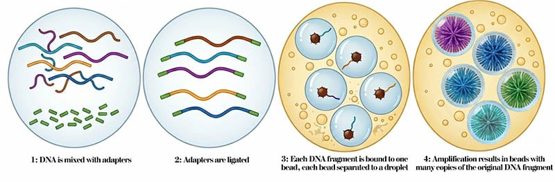
Figure 1. ePCR workflow, step by step. Adaptors are ligated to fragments, bound to beads, isolated in droplets, and amplification results in multiple copies of the fragment, with each droplet having an enriched single sequence.
Both Ion Torrent and 454 pyrosequencing used sequencing by synthesis (SBS). In each case, amplified DNA was attached to beads and loaded into microplate wells (1 bead/well). Nucleotides were then flowed in one at a time, and incorporation was detected by a signal change. In 454 pyrosequencing, correct nucleotide incorporation released pyrophosphate (PPi), which was converted to ATP and then to light by luciferase, and the light was measured by a camera. In Ion Torrent, incorporation was detected by the release of hydrogen ions, which changed pH and was measured by a semiconductor sensor.
Early sequencers required separated fragments with enough enrichment to generate reliable signals. ePCR solved these issues but also added workflow complexity. Regardless, 454 pyrosequencing and Ion Torrent both thrived thanks to ePCR, which was the key enabling step for the first wave of commercial NGS.
Bridge amplification and Illumina
Illumina launched its first commercial sequencer, the Solexa 1G, in 2006. It already used the two processes it is well known for: bridge amplification coupled with SBS. Here is a rundown of bridge amplification, which creates clustered copies of DNA fragments on a glass flow cell:
- Illumina adapters are ligated to the fragmented DNA.
- The molecules are loaded into a single flow cell with many primers complementary to adapters attached to it.
- The DNA fragments bend so both adapters bind to oligos in the flow cell, creating “bridges”.
- The fragments are amplified.
- Heat is used to denature the dsDNA, and after cooling, each strand will form another bridge hybridizing with another primer.
- The process is repeated, resulting in each individual fragment creating a small cluster of sequences all attached next to each other.
After amplification, all of the reverse strands are cleaved and removed. Complementary strands to the forward strands left are polymerized, with fluorescently tagged dNTPs added one by one. The fluorescent color of the dNTP added is recorded. Different colors are used for different dNTPs, which makes it possible to determine the sequence of the synthesized fragment.
Bridge amplification is tightly coupled to Illumina because the flow cell, surface oligos, and fluorescence SBS are all designed as one integrated system.
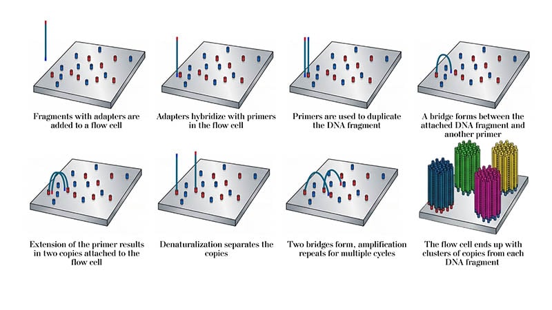
Figure 2. The process of bridge amplification
Rolling-circle amplification and DNBSEQ
Another type of amplification, called DNA nanoball (DNB), was developed for the Complete Genomics platform. This approach leverages phi29 DNA polymerase’s high processivity and strand displacement capabilities to create long DNA products that self-fold into balls or dense molecules from circular DNA templates. The process goes as follows:
- Fragment DNA if needed and add adapters.
- Circularize the library and digest leftover linear DNA.
- Rolling-circle replication/amplification is used to generate a DNA nanoball. Phi29 DNA polymerase is used to extend primers going around the circular templates many times. Its strand displacement quality allows it to unwind dsDNA, its high processivity results in extra-long products, and the 3’–5’ exonuclease activity corrects errors, which allows for better coverage than PCR.
- The produced ssDNA products self-assemble into clusters or nanoballs thanks to self-complementary adaptor regions.
- The nanoballs are loaded into high-density patterned flow cells for sequencing by synthesis.
DNB sequencing (DNBSEQ) uses linear amplification but results in high DNA density. The sequencing is similar to Illumina, but the clusters are DNB’s instead of bridge amplification.
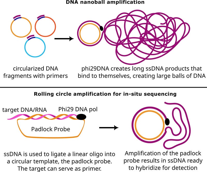
Figure 3. DNB and RCA explained. Both types of amplification rely on circular templates amplified with phi29 DNA polymerase.
DNA amplification in fourth-generation sequencing
Ion Torrent, 454, Illumina and DNBSEQ are part of the second-generation sequencing wave: nucleic acids are extracted, amplified, and sequenced. Third-generation sequencing generally refers to single-molecule sequencing, with no amplification required, using platforms like PacBio and Oxford Nanopore, which allow detection of markers in the original nucleic acid sequence (such as epigenetic markers) and longer reads. But fourth-generation sequencing deals with spatial resolution, and often uses amplification to achieve enough copies to be detected.
A common amplification format is rolling circle amplification (RCA) with padlock-probes, used for in-situ sequencing (ISS). Here’s how it works:
- Fix and permeabilize the cells or tissue so reagents can access the targets while keeping spatial structure intact.
- Add gene-specific padlock probes or oligos that hybridize to target RNA or cDNA-derived sequences in place.
- Ligate the correctly matched probes to form circular templates only at the target site.
- Introduce phi29 DNA polymerase and other amplification reagents, usually in a solution over the tissue section.
- Perform RCA, letting phi29 DNA polymerase create long ssDNA products, to generate a spatially confined DNA concatemer.
- The products can be detected using sequencing-by-hybridization / sequencing-by-ligation cycles.
- Because the product stays anchored where the target originally was, the resulting signal preserves the tissue’s spatial context.
Some instruments have been developed to read ISS, and others use simple fluorescence microscopy or other systems, but the field of fourth-generation sequencing is still developing.
Regardless, amplification of DNA/RNA is used for many types of sequencing. Understanding how amplification works for any given workflow, which biases it introduces in sequencing, and how they may affect your samples is crucial to improve your sequencing data and experiments.