Multiplexed imaging enables the visualization and quantification of many different biomarkers within the same tissue sample, offering valuable insights into how the cells expressing those biomarkers are interacting. Here, we look at the changing landscape of fluorescence-based multiplexed imaging and suggest factors to consider when selecting a suitable platform for your research.
The evolution of fluorescence-based multiplexed imaging
Broadly speaking, fluorescence-based techniques for multiplexed imaging of proteins and mRNA use fluorophore-labeled antibodies (immunofluorescence) and fluorophore-labeled complementary oligonucleotide probes (fluorescence in situ hybridization) to detect targets of interest, respectively.
Search Spatial biology platforms Search Now Search our directory to find the right spatial biology platform for your research needs.
Immunofluorescence was first demonstrated in 1941, when Coons et al. used a fluorescein isothiocyanate (FITC) labeled antibody to visualize Pneumococcal bacteria in infected murine tissues. Following this, advances in antibody labeling technology soon made it commonplace for researchers to simultaneously detect 2–4 different markers—a number limited by the need to avoid spectral overlap. More recently, the development of iterative staining methods that involve cycles of antibody incubation followed by dye inactivation/antibody elution has allowed for imaging tens of different markers in formalin-fixed, paraffin-embedded (FFPE) sections, often with the aid of automation.
One of the most quantitative techniques for mRNA detection is single molecule fluorescence in situ hybridization (smFISH). This involves hybridizing large numbers of short, fluorescently labeled oligonucleotide probes to specific targets in FFPE sections such that each mRNA molecule can be visualized and quantified via fluorescence microscopy. To increase the number of targets detected, as well as improve experimental accuracy, smFISH has evolved into approaches including multiplexed error-robust FISH (MERFISH), which can measure the copy number and spatial distribution of tens of thousands of RNA species in individual cells, using binary barcodes to detect and correct errors.1-3
Key impact areas
A main reason for researchers to choose higher-plex imaging techniques, such as iterative immunofluorescence and MERFISH, is that these methods allow for visualizing a broader diversity of cell types, functional states, and interactions within the same tissue sample than traditional approaches where only a small subset of the cells are characterized.
“Areas that are especially benefiting from advances in multiplexed imaging include cancer research, where scientists can now better map tumor microenvironments, monitor disease progression, and understand how immune cells and cancer cells interact,” explains George Emanuel, Ph.D., Co-founder and VP of Instrumentation at Vizgen, Inc. “Additionally, multiplexed imaging has seen widespread uptake for neuroscience research, where it is used to distinguish neural cell types, map brain circuits, and investigate the molecular changes underlying neurodegenerative disorders such as Alzheimer’s disease.”
Other key impact areas include immunology, where multiplexed imaging enables the analysis of immune cell distribution and activation in autoimmune diseases and infections, and regenerative medicine, where multiplexed imaging is used for studying stem cell differentiation and tracking tissue repair. “Multiplexed imaging has also proven useful for pharmacology and drug development, not only for evaluating the efficacy of potential drug candidates, but also for detecting any unintended impacts, such as toxicity,” says Emanuel.
Proteomic and transcriptomic imaging technologies
While there is a wide variety of fluorescence-based multiplexed imaging technologies, the following are leading examples of platforms with both proteomic and transcriptomic imaging capabilities.
• Cell DIVE multiplexed imaging solution
Cell DIVE, available from Leica Microsystems, allows researchers to detect more than 60 different protein biomarkers on a single FFPE tissue section through an iterative staining and dye inactivation workflow. “Cell DIVE is an advanced, open multiplexed imaging platform that enables high-plex, whole-slide imaging and single-cell analysis of tissue samples,” reports Betty Yuen-Torres, Global Marketing Manager Spatial Biology. “With Cell DIVE, researchers can choose which antibodies they use—including the option to select from 350+ Cell DIVE validated antibodies—and the level of automation they require.”
The Cell DIVE acquisition software now supports Z-stack imaging for detailed visualization of cellular structures. “In combination with Aivia AI-powered image analysis software, Cell DIVE allows for quantifying protein expression and assessing cellular morphology to reveal complex spatial and molecular patterns,” says Yuen-Torres. “In addition, there is the option for multi-omics workflow integration, including automated RNA staining and imaging and multi-round acquisition using the BioAssemblyBot® 200—a customized multi-tasking robot that seamlessly integrates with the Cell DIVE imager.”
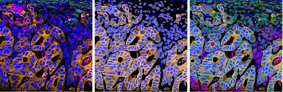
Figure 1. 2D slice of colon cancer tissue stained with 30 markers and imaged using the Cell DIVE system. Analysis performed using Aivia’s multiplex cell detection recipe and automatic clustering tool. Original image (left), detected cell membrane shown in white (middle), each phenotype denoted in a different color (right). Images provided by Leica Microsystems.
• MERSCOPE Ultra™ Spatial Imaging
MERSCOPE Ultra™ Spatial Imaging, available from Vizgen, offers a tissue-wide view of up to 1,000 custom genes at single-cell resolution. Based on MERFISH technology, it captures spatially resolved transcriptomic data at sub-cellular resolution across large tissue sections—up to 3 cm2 and 3 million cells on a single slide. “By incorporating error-robustness through binary barcoding of RNA, MERFISH ensures highly accurate and highly quantitative measurement of RNA transcripts to reveal what’s happening within all the cells across the whole tissue with fine detail,” reports Emanuel. “Moreover, with the launch of MERFISH 2.0 earlier this year, researchers can now achieve even greater sensitivity, including from archival or clinical samples, in which the RNA is often more degraded.”
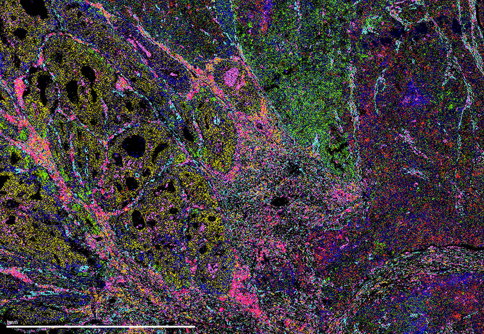
Figure 2. Human uterine cancer sample. Image provided by Vizgen.
For those wishing to integrate transcriptomics with proteomics measurements, MERSCOPE currently allows users to detect up to 9 custom proteins using oligo-conjugated antibodies. This number is set to increase to around 20–30 in the near future, with continued development of the underlying technology.
Future perspectives
While identifying a handful of cell types in FFPE tissue was once considered sufficient, there is a growing trend toward obtaining more nuanced findings. Fluorescence-based multiplexed imaging platforms are delivering on this need, helping to answer questions such as which cell types interact under conditions of disease, which signaling pathways influence disease pathology, and how does aberrant signaling alter cellular function. Using platforms such as those discussed here, researchers can take spatially resolved single-cell resolution to new levels.
Factors to consider when selecting a fluorescence-based multiplexed imaging platform
- Do you want to detect proteins, mRNA, or both?
- Can you customize target detection?
- What sort of throughput do you need?
- What level of sensitivity do you require?
- Is tissue preservation important?
- Is the platform easy to use, including compatibility with automation?
- Can the platform keep up with the evolution of your study?
References
1. Chen KH, Boettiger AN, Moffitt JR, et al. RNA imaging. Spatially resolved, highly multiplexed RNA profiling in single cells. Science. 2015;348(6233):aaa6090.
2. Moffitt JR, Bambah-Mukku D, Eichhorn SW, et al. Molecular, spatial, and functional single-cell profiling of the hypothalamic preoptic region. Science. 2018;362(6416):eaau5324. doi:10.1126/science.aau5324
3. Cohen L, Halpern A, Blosser TR, et al. Whole-transcriptome-scale and isoform-resolved spatial imaging of single cells in complex tissues. bioRxiv preprint