Solid tumors can be considered to be “sensitive” or “resistant” depending on their resistance to a class of therapeutics called immune checkpoint inhibitors (ICI), generally demonstrating a durable and significant response to this treatment approach in only about 20% of all cancers. This response can be linked to the tumor itself, its surrounding microenvironment (TME), as well as the presence of a variety of cell types in addition to the cancerous cells, including fibroblasts and endothelial cells. The immune cells that often penetrate tumors have been shown to be particularly fundamental to such a response, including effector T cells, T lymphocytes, tumor-associated macrophages, dendritic cells, and natural killer cells.
Search Spatial biology platforms Search Now Search our directory to find the right spatial biology platform for your research needs.
This article looks at how immunophenotyping tumors as “hot” and “cold” based on their immune cell penetration can provide a useful indicator of tumor response to therapeutic strategies. It also discusses current methodologies and their potential for future translation in the clinic.
Understanding hot and cold tumors
A low density of tumor-infiltrating T cells can contribute to ICI resistance, with those specifically low in their density of tumor-infiltrating T lymphocytes called cold. Those that are hot demonstrate more dense effector T-cell infiltration and often are significantly more responsive to ICIs. Tumor biopsies can provide insight into the immune cell TME as well as tumor mutational burden and can be used to pinpoint patients who might have more of a response to immunotherapeutic approaches.
Natasha Lewis, Senior Scientist on Sartorius’s BioAnalytics Applications Team, comments that “the classification of tumors as either hot or cold has become a major paradigm in the cancer field. This is so particularly in the development of immunotherapies relying on T cells that aim to identify, then eliminate, cancer cells.”
“There are two main questions,” explains Anastasiia Marchuk, Junior Product Specialist at TissueGnostics: “1) What exactly dictates if the tumor becomes hot or cold, and 2) What can we do to turn an unresponsive cold tumor into a responsive hot tumor?” Applying tumor immunophenotyping can help with answering such questions.
From tumor immunophenotyping to precision medicine
Tumor immunophenotyping, or classifying cells of the immune system based on different T-lymphocyte subsets, for example, by the presence or absence of surface antigens, is an important area of research. It has the potential to improve the efficacy of immunotherapies and give rise to precision medicine. “Ultimately, this [approach] will allow for better patient selection for current treatments and also identification of novel targets for new treatments, including combinations,” says Subham Basu, Chief Business Officer, Navinci Diagnostics.
Lewis shares her perspective on the translational potential of tumor immunophenotyping approaches: “One of the obvious translational applications of immunophenotyping is the potential for personalized medicine based on predictive biomarkers, to select the most appropriate treatment regime for each patient and reduce trial-and-error approaches.” If such personalized medicine approaches are realized in the future, it could have marked benefits for patients and clinicians in terms of both time and cost.
Immunophenotyping is commonly carried out via flow cytometry. Unfortunately, the flow approach does not provide a three-dimensional view of the tumor and wider TME composition in terms of how those aspects might influence tumor responsiveness to ICIs. Marchuk comments: “Flow cytometry was used as a method of choice for decades because it allowed fast detection and sorting of cells based on the specific proteins they express. Today, it is not enough to only know which cells and how much of them are present in the sample—we need to know how they are distributed. This need has led to a rise in spatial biology approaches.”
Traditional immunohistochemistry (IHC) or immunofluorescence (IF) staining, when combined with whole-slide imaging, can also help better understand the distribution of particular cell types in their native tissue microenvironment. Marchuk describes a recent study combining spatial transcriptomics and multiplex immunofluorescence imaging to characterize subsets of tumor-associated macrophages (TAMs) known as monocyte-derived TAMs in glioblastoma. The research group used TissueGnostic’s TissueFAXS Spectra for whole-slide multispectral imaging and StrataQuest image analysis solution.
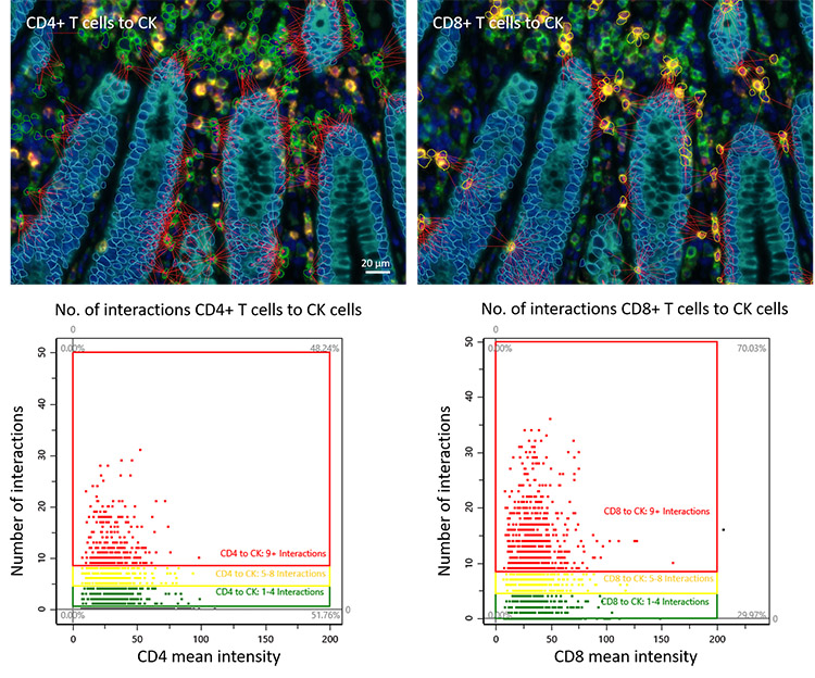
Phenotype interaction analysis of CD4+ and CD8+ T cells with CK+ cells in colorectal cancer in StrataQuest. Image courtesy of TissueGnostics.
Tissue cytometry allows for the study of cells and their interactions over the whole tissue section, which allows the investigation of the TME. The quantity of particular cell phenotypes, their distribution, or proximity to certain structures within the TME, can indicate a biological process directly influencing disease progression.
Newer methods
“Our technology,” states Basu, “allows for detection, visualization, and analysis (including quantification) of protein-protein interactions, whether they’re between cells (such as receptor-ligand interactions, e.g., PD-1, PD-L1) or intracellular signalling (e.g., PD-1, SHP-2). Both allow for actual interrogation of protein function in the TME between cell types (e.g., tumor-immune, immune-immune, tumor-fibroblasts).” Understanding TME protein function can facilitate the monitoring of drug mechanism of action, evaluation of mutational background, differences in cell type make-up, detection of novel drug targets, and selection of biomarkers, as well as basic research in signalling pathways.
Multiple validated assays also deliver high-quality data on cell behavior, including migration and invasion, immune cell infiltration, and apoptosis in response to treatments. Lewis comments: “Fluorescent reporters and reagents can be used to monitor the dynamics of signaling pathways in real-time in response to treatments. Real-time monitoring of cellular dynamics using Sartorius’ Incucyte® can be very powerful for dissecting the TME. The iQue® High-Throughput Screening Cytometry Platform allows for rapid and simultaneous analysis of multiple parameters at the single-cell level. And bead-based assays for the iQue® to quantify chemokines and cytokines can characterize tumors' inflammatory states, how they change throughout tumor responses, or when an inflammatory response has been initiated.”
In terms of even newer methods on the horizon, researchers are also investigating methodologies that help monitor dynamic changes in the TME, with deeper sequencing and improved proteomic methodologies for antigens being explored.
“Mass cytometry (cytometry by time of flight, or CyTOF) allows measurements of up to 50 target surface markers, although acquisition can be up to an order of magnitude slower than using flow cytometry,” states Lewis. “High dimensional flow cytometry allowing a larger panel of markers can provide a more detailed phenotypic profile of immune cells.” At the moment, advanced computational tools and machine learning algorithms are also in development in order to handle the large and complex datasets generated by such new immunophenotyping techniques, with improved algorithms for data integration, normalization, and visualization.
The road to 3D and AI
What does the future hold for immunophenotyping hot and cold tumors? “The use of 3D models in research and drug development also has huge potential and opportunity. These models are more predictive than a 2D culture or a mouse model that may not replicate a human tumor sufficiently and may reduce drug failure in clinical trials, saving both time and money,” predicts Lewis. Using iPSCs in such culture systems could also potentially lead to advanced personalized treatment strategies.
Basu and the team at Navinci feel that the next set of advances “will come from greater exploration of protein function, not just a greater ‘plexy’ with multiplex immunofluorescence or deeper next-generation sequencing.” Currently, the company’s assays are research-use only but have been used to enrich biomarker evaluation in retrospective analysis of clinical trials in immuno-oncology studies, including for non-small-cell lung cancer, hereditary nonpolyposis colorectal cancer, colorectal cancer, and bladder cancer.
“Surely, as we accumulate more knowledge about how our immune system combats cancer, there are even more questions to be answered,” concludes Marchuk. “Artificial intelligence is already a new standard, especially in image analysis—helping to handle large volumes of data, analyzing it, and ensuring efficiency and accuracy. That’s why we definitely expect to see more and more examples of it being implemented in workflows in the future.”
References
1. Zhang J, Huang D, Saw PE, Song E. Turning cold tumors hot: from molecular mechanisms to clinical applications. Trends Immunol. 2022 Jul;43(7):523-545.
2. Zabransky DJ, Yarchoan M, and Jaffee EM. (2023). Strategies for heating up cold tumors to boost immunotherapies. Annu Rev Cancer Biol. 2023; 7:149-170.
3. Wang W, Li T, Cheng Y, et al. Identification of hypoxic macrophages in glioblastoma with therapeutic potential for vasculature normalization. Cancer Cell. 2024;42(5):815-832.e12.