Cytokines are essential cell signaling molecules with links to conditions including Alzheimer’s disease, rheumatoid arthritis, and cancer. This article looks at current methods for detecting and measuring cytokines and comments on how these are being used.
Why study cytokines?
Cytokines have long been known to modulate the development, activation, and maturation of immune cells, typically via autocrine (on the same cell) or paracrine (on a neighboring cell) action. However, cytokines have also been implicated in multiple disease states, making them important molecules for therapeutic targeting. “Cytokine levels change after tissue injury, inflammation, and in response to acute and chronic infection, as well as during sepsis, autoimmune disease, and cancer,” reports Paul Wylie, Ph.D., Director for Multiplex immunoassays at Abcam. “In addition, physiological processes like aging are associated with altered cytokine expression, while the over secretion of cytokines has recently been observed as a cause of deaths during the COVID-19 pandemic and various immunotherapy trials. For these reasons, being able to accurately detect and measure cytokines is crucial to understand how they function and improve the lives of patients.”
Complicating factors
A main challenge when analyzing cytokines lies in understanding how they act. While some cytokines are redundant (share the same functionality as other cytokines), others are pleiotropic (have different effects on different cell types), synergic (require that other cytokines be present in order to exert their function), or antagonistic (inhibit one another), which must always be considered when drawing conclusions from experimental data.
Cytokine multiplex assays Search Now Search our directory to find cytokine multiplex assays for your research needs.
Beyond this, several further properties of cytokines make studying them difficult. “Cytokines are often expressed at ultralow concentrations—into the picomolar range—meaning highly sensitive immunoassays are required for their detection,” explains Anne Sloan, Ph.D., Technical Scientist at Cell Sciences. “They are also extremely dynamic, having short half-lives due to the transient nature of most cytokine secretion processes, and can exhibit very localized intracellular signaling.”
Establishing normal cytokine levels can also present problems. “When it comes to data analysis, there is high interindividual variability in cytokine levels and level changes,” explains Laurence Loi, Product Manager for Immunoassays at Abcam. “Researchers must therefore establish ‘normal’ versus abnormal levels and accurately determine how changes can be attributed to disease pathogenesis. Fortunately, suppliers have been working on improving the accuracy and sensitivity of their assays to help with this.”
Is one cytokine enough?
“As cytokine research has evolved, researchers have learned the importance of characterizing cytokines both as individual biomarkers and in the context of a coordinated response,” says Anna Nikolenko, Ph.D., Product Manager for Immunoassays at BioLegend.
“For example, while IL-6 is now known to have a critical role in the development and progression of autoimmune diseases including rheumatoid arthritis, and IL-2, IL-7, and IL-15 have all demonstrated therapeutic potential as cancer immunotherapies, it is common to find a strong relationship between multiple cytokines. This makes multiplex immunoassay panels that target several cytokines related to a specific disease or condition extremely useful.”
Sloan adds that it is common for panels to comprise pro-inflammatory (e.g., IL-1β, IL-6, IL-8, IL-12, TNF-α, and interferons) or anti-inflammatory (e.g., IL-4, IL-6, IL-10, IL-11, IL-13, IL-1 receptor antagonist, and TGF-β) cytokines, which she says can be used to help unravel the etiology of infectious or chronic inflammatory disease. For example, panels of proinflammatory cytokines have been widely used to investigate the cytokine storm associated with COVID-19. Yet she cautions that a single cytokine may have pro-inflammatory or anti-inflammatory properties depending on context. “Irrespective of whether you’re using a panel or analyzing an individual cytokine, finding out as much as possible about the biology of your target before undertaking any research is key,” she says.
Methods for cytokine detection and analysis
Immunoassays are by far the most popular approach to studying cytokines, with ELISA, ELISpot, and intracellular flow cytometry leading the way. Broadly speaking, ELISA is used for measuring cytokines in liquid samples such as serum, plasma, or cell culture supernatant; ELISpot for determining the frequency of cytokine-secreting cells following their introduction to an antibody-coated surface; and intracellular flow cytometry for identifying which cell types secrete different cytokines. In addition, Luminex Corporation’s xMAP technology, which uses a bead-based approach for detecting as many as 500 analytes in a single sample, is widely literature cited for cytokine research and has recently been augmented with the launch of xMAP INTELLIFLEX®, which can simultaneously acquire two parameters per analyte.
“The advantages of multiplexing are that you can obtain more data per sample, while also realizing labor cost savings by not having to run a separate assay for each individual analyte,” notes Wylie. “Our FirePlex® platform allows researchers to quantify multiple cytokines per sample with a flow cytometer or high content imager, using the same recombinant antibody pairs as our SimpleStep ELISA® kits—90-minute single-wash sandwich ELISAs that cover all major cytokines in human, mouse, and rat samples. The individual antibody components can also be purchased separately for consistency across different assay formats.” Loi adds that published applications of these technologies include the use of FirePlex for measuring cytokine release during the large-scale production of human iPSC-derived macrophages for drug screening, and the measurement of inflammatory cytokines by SimpleStep ELISA within a study to investigate the relationship between circulating BMP4 and perioperative inflammation.
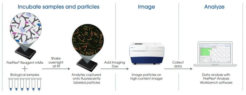
FirePlex assay workflow. To capture analytes onto FirePlex particles, biological samples are added to a 384-well imaging plate and incubated with the FirePlex-384 Reagent Mix overnight. Subsequently, an imaging dye is added, and plates are scanned on high-content imagers. The FirePlex Analysis Workbench software generates standard curves and quantifies analytes of interest directly from image files. Image provided by Abcam.
BioLegend’s cytokine products include a bead-based system known as LEGENDplex™, which uses the same basic principles as sandwich immunoassays to enable multiplexed research. “This year, BioLegend was declared a ‘Supplier to Watch’ for having the largest percentage increase in citations for multiplex products,” reports Nikolenko. “Indeed, over the years, a growing number of publications have referenced our LEGENDplex panels for cytokine research. For example, in 2018, our LEGENDplex™ Human Th Panel was cited by Yamaguchi et al. for measuring the cytokine concentration in samples obtained from patients undergoing treatment with nivolumab, an anti-PD-1 therapy, where it contributed to the discovery that nivolumab exerts its effects through activation of central/effector memory T cells and T‐helper 1 polarization. More recently, the same product was referenced in a publication demonstrating the importance of T cells in SARS-CoV-2 immunity, where it was used to characterize their cytokine response to the virus.”
A bright future
With cytokines being fundamental to such a diverse array of cellular processes, they remain the focus of research groups worldwide. Suppliers such as Abcam, BioLegend, and Cell Sciences are supporting these efforts by developing novel products for cytokine detection and analysis, helping ensure that the number of approved cytokine therapies continues to grow.
Doubling the number of readouts per sample
Luminex’ xMAP technology uses color-coded beads that are pre-coated with antibodies to bind up to 500 different analytes in solution. Analyte capture is detected by adding biotinylated target-specific antibodies and PE-conjugated streptavidin before reading on a dual-laser flow-based instrument, where one laser (635 nm) identifies the bead and the other (532 nm) measures the magnitude of the PE-derived signal. xMAP INTELLIFLEX® builds on the original xMAP technology by incorporating a second (405 nm) detection channel. This enables simultaneous measurement of two parameters per analyte on a single bead, effectively doubling the number of readouts obtained per sample. For example, using xMAP INTELLIFLEX® for cytokine analysis, it is possible to both detect a target of interest and track a specific post-translational modification.