Although there are still some particles too small to detect using flow cytometry, modern advances have made it possible for researchers to interrogate sub-micron entities such as extracellular vesicles (EVs), viruses, and subcellular organelles in statistically relevant numbers. In this article, we explain what’s involved in nanoscale flow cytometry and provide guidance for getting the best out of single nanoparticle analysis applications.
Nanoscale particle detection presents unique challenges
The main difference between conventional flow cytometry and nanoscale flow cytometry lies in the size of the analyte. “Common flow cytometry analytes like cells and bacteria have a diameter greater than 1 µm, with human white blood cells typically ranging from 10–20 µm,” explains Pia Jeggle, Ph.D., group leader product management, flow cytometry instrumentation at Miltenyi Biotec. “In contrast, nanoscale flow cytometry analytes have a diameter from ≤1 µm down to as little as ~50 nm and vary considerably in terms of their composition and corresponding refractive index. This makes detecting them with flow cytometers designed for applications like cell immunophenotyping challenging.”
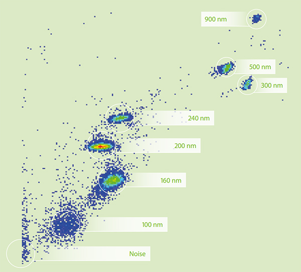
Image: Using the violet side scatter optics of the MACSQuant® Analyzer 16 allows for small particle analysis. Image provided by Miltenyi Biotec.
According to Lauren Jachimowicz, applications development scientist at Agilent Technologies, a major difficulty when working with nanoscale particles involves discriminating them from any background signal. “Nanoscale particles are more difficult to resolve from light scatter compared to cells,” she says. “Additionally, the resolution of surface markers on nanoscale particles is more challenging due to the small size. For example, a 100 nm nanoparticle will have 10,000 times fewer surface molecules and 1 million times fewer cellular contents than a 10 µm cell. Accurately detecting such a particle therefore requires an extremely sensitive flow cytometer.”
Modern instrumentation enables novel discoveries
Fortunately, flow cytometry instrumentation has evolved to meet the demands of nanoscale particle detection. “Some cytometers are optimized to measure biological particles down to 100 nm by light scatter and can also measure weak fluorescence,” reports Oliver Kenyon, CEO at Apogee Flow Systems. “These capabilities have driven rapid growth in many novel areas, including the emerging use of extracellular vesicles as biomarkers for therapeutic and diagnostic applications.”
Fanuel Messaggio, senior development scientist, flow reagent R&D team at Beckman Coulter Life Sciences, comments that EV characterization is probably the top application for nanoscale flow cytometry at present. “EVs represent a physiological mechanism of communication between cells,” she says. “They can be released by every cell type and are found in every biofluid, making their investigation by flow cytometry a priority for many research groups. In the last decade, EV publications have increased 10-fold, and there has recently been a strong focus on standardizing their flow cytometric detection and analysis.”
Importantly, studying EVs and other nanoparticles does not necessarily require a dedicated flow cytometry instrument. Instead, conventional flow cytometers can often be adapted for nanoscale particle detection. “Instruments such as the ZE5 Cell Analyzer have small particle upgrades that allow the 405 nm laser to achieve the sensitivity needed for nanoscale flow cytometry,” notes Yasha Talaga, applications and collaborations manager, Bio-Rad Laboratories. “The paper held up by Dr. Fauci in a SARS-CoV-2 press conference cited work done at La Jolla Institute on a ZE5 Cell Analyzer, highlighting virus research as another major driving factor in the uptake of nanoscale flow cytometry. To put this into further context, in 2013, studying viruses by flow cytometry had increased to such a point that the term ‘flow virometry’ was coined.”
Other notable developments in the instrumentation used for nanoscale flow cytometry include Apogee Flow Systems’ Micro-PLUS (which also offers particle sorting) and Beckman Coulter’s CytoFLEX, for both of which the smallest detectable particle size is defined using polystyrene beads. These are complemented by Miltenyi Biotec’s MACSQuant® Analyzer 16 that is equipped with violet side scatter optics for nanoparticle analysis and Agilent’s NovoCyte Quanteon that combines two modern detection technologies. “Avalanche photodiodes enable the detection of side scatter, while silicon photomultipliers (SiPM) are used for highly sensitive fluorescent and light scatter detection,” reports Jachimowicz. “But it is important to remember that whichever instrument you have access to, you should always confirm it is sufficiently sensitive for nanoparticle analysis before initiating any experiments.”
Key considerations for performing nanoscale flow cytometry
The detection limits of your flow cytometer can readily be determined using small particle and dim fluorescence beads, yet many other factors besides the sensitivity must also be considered. “When it comes to nanoscale flow cytometry, sample prep makes all the difference,” says Talaga. “Depending on the particle you are studying, media should be filtered, potentially de-gassed, and appropriate for your sample type. Using small, bright fluorescent dyes can increase your chances of target detection. And knowing the approximate size of your nanoparticle is important in instrument setup as well.”
“Controls are essential for small particle analysis,” adds Jachimowicz. “Setting up a dilution curve can determine if any coincidence (where two particles pass the laser at the same time and are qualified as one event) is occurring. You should see a linear decrease in event concentration with no change to the mean fluorescent intensity of the sample. It is also advised to include a control where the membranous vesicles have been destroyed with detergent (e.g. by treating the sample with 1% Triton-X). This ensures you are detecting your particle of interest and not aggregates of antibodies, dyes, or other substances.”
Jeggle also points out the importance of the observation volume, a parameter she feels is not often discussed in flow cytometry. “The observation volume is based on the angle of the incident and detection light in combination with the height of the excitation light,” she explains. “If you imagine that the observation volume is in the range of ~25x25x10 µm, then an average white blood cell with a diameter of 10 µm would occupy about a tenth of this, making it easily distinguishable from the background. However, a particle with a diameter of ~200 nm would occupy only a millionth of the observation volume, meaning that small disturbances and impurities could prevent it from being clearly identified. For this reason, and because the likelihood of coincidence is also much greater for small particles, the observation volume should always be kept as small as possible.”
Lastly, Maria C. Gentile, senior manager, product management, biodiscovery hardware at Beckman Coulter Life Sciences, mentions the value of clean-up methods. “Size exclusion, affinity, and ion exchange chromatography can be used to enrich your (EV) target or deplete non-target particles,” she says. “By following MISEV and MIFlowCyt-EV guidelines when employing experimental strategies for sample preparation and characterization as well as instrumentation setup, you will improve the likelihood of generating high-quality data that you can share with confidence.”