Since nerves control everything that happens in our bodies, neuroscience has become a treasure trove of discoveries fueling basic science, medicine, and the all-encompassing activity known as drug development. Of prevailing interest are neural events with significant characteristics in the molecular, spatial, and time domains, and observed in both cultured cells and living organisms.
In other words: Microscopy to the rescue!
Advances in microscopy now permit simultaneous visualization of critical processes in all three of those domains. Even within the physical constraints imposed on optics (e.g. there will never be an optical transistor), what continues to amaze regarding the marriage of microscopy and nerve cells is the adaptability of older microscopy methods to today's scientific questions.
Oldie but goodie
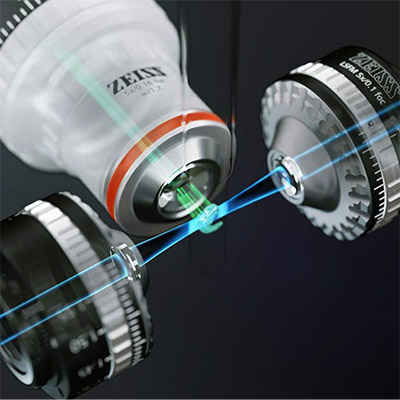
Light sheet fluorescence microscopy (LSFM), a planar illumination technique for imaging biological specimens, has been known for more than 100 years but its first practical application was reported in 1993. LSFM splits fluorescence excitation and detection into two separate, perpendicular light paths. This allows illumination of a thin section of the sample without filtering, pinholes, or image processing, while optimizing photon collection from the region of interest. Orthogonality between illumination and detection maximizes detection of the target region by minimizing the effects of fluorescence from out-of-focus features.
By combining optical sectioning with parallel image acquisition from the focal plane, LSFM allows image collections more rapidly than with confocal microscopy, and with less excitation light. That means experiments can proceed for days without photobleaching—a boon for imaging live nerve cells.
ZEISS claims its Lightsheet 7 LSFM incorporates dedicated, adjustable optics, sample chambers, and sample holders that enable imaging of optically cleared specimens, as large as mouse brains, at subcellular resolution.

For more detail on LSFM, including manufacturers, check out our recent review article.
Optical clearing is required for large biological specimens due to sample inhomogeneities. Courtney Akitake, Ph.D., Product Marketing Manager at Zeiss, explains that thick samples tend to scatter light omnidirectionally. "Light entering the sample traveling in one direction ends up going off into all different directions. Different components of biological tissues such as lipid, protein, and intra- and extracellular fluids, all have different refractive indices, and scatter is related to refractive index transitions as light passes through a heterogeneous sample. Clearing corrects for these heterogeneities while maintaining 3D structure. After clearing, light travels mostly along the same direction that it entered, allowing deeper, non-invasive imaging without physical sectioning."
LSFM allows experimenters to visualize the entire organ, e.g. mouse brain, and then probe deeper into the tissue or cell. A major advantage of imaging at both low and high magnification is that it facilitates finding a region of interest in large cleared tissues. "With Lightsheet 7, researchers can acquire an overview of a whole cleared mouse brain and then efficiently navigate to a region of interest to image axonal projections of individual neurons," Akitake says.
Neuroscience may have cut its eyeteeth on the study of fixed specimens, but modern science is about probing dynamic, i.e. living, systems. Where imaging fixed specimens provides a snapshot of the sample frozen in time, LSFM allows real-time investigations into neurogenesis, axonal guidance in development, and functional calcium imaging—all in living systems. "The method's sensitivity, speed, and integrated environmental controls creates ideal conditions for living organisms, allowing researchers to study dynamic processes over hours or even days," Akitake tells Biocompare.
No one-hit wonder
Intact tissue and cultured neurons are both common sample types in neuroscience, but since sample material is usually in very short supply, investigators must design exquisitely efficient assays to get the most from their samples. Confocal microscopes have long been a staple instrument for acquiring three-dimensional images. While conventional imaging experiments have been limited to three- or four-color imaging, confocal microscopes are now capable of as many as 32 colors or wavelengths.
"Sample type and analytical needs determine which type of microscopy is best suited for a particular experiment," says Rebecca Bonfig, Product Manager at Olympus Life Science.
Bonfig explains that, for example, a culture of primary neurons with one or two spectrally distinct labels may best be observed on a widefield epifluorescence or spinning disk microscope, whereas a thicker brain section with three or four colors is better suited for a point-scanning confocal microscope. "For even thicker intact tissue samples, such as whole cleared brains or in vivo brain imaging, a multiphoton microscope is the best solution, as the longer wavelength near-IR excitation light can reach much deeper into a tissue sample with less scattering than visible light."
In microscopy, the number of colors/wavelengths approximately tracks with the number of individual questions investigators can expect to answer from an experiment. With spectral unmixing technology, a technique for deconvolving overlap in fluorescence emission bands, confocal microscopes can detect anywhere from 16 to 32 individual channels. "In practice, however, the limitation typically comes from the number of probes that you can reasonably 'fit' into a sample without running into spectral crosstalk," Bonfig adds.
The development of robust fluorophores across the spectrum, and especially into the red and near-infrared regions, has made five- and six-color experiments easily achievable while maintaining good color separation between channels.
One technique that catapulted multiplexing experiments and vastly expanded the number of colors that can be imaged in a sample is Brainbow imaging, which relies on the random expression of three different fluorescent proteins at various levels in individual neurons. With a different ratio of blue, green, and red fluorophores occurring in each neuron, the Brainbow technique generates more than 100 different colors, thus enabling researchers to easily distinguish individual neurons from neighboring dendritic and axonal processes. "Brainbow has been transformative for microscopy applications in neuroscience, including brain mapping and neural circuit studies that provide insight into neurological and psychological disorders and potential targets for treatment," says Bonfig.
One of the most straightforward but under-appreciated approaches to getting more information from samples is spectral or “lambda” scanning, which creates a spectral profile of all signals present in a sample. "Similar to how a Z-stack captures an image by stepping through Z positions one at a time, lambda scans work by stepping through the emission spectrum in discreet, predefined steps," Bonfig explains. "Just as users can define how small or large of a Z step to take, they can assign a detection bandwidth and step sizes in a lambda scan, depending on the labels present." For example, they can designate larger bandwidths and step sizes for samples with spectrally distinct fluorophores, whereas a sample with highly overlapping fluorophore emission spectra will benefit from smaller detection windows.