The ability to accurately assess cell proliferation is critical to research at all stages and across fields, especially immunology and oncology. Cell proliferation assays are used to evaluate normal cell health, measure responses to drugs, or as a prognostic and diagnostic tool in cancer. There are a number of methods that vary with respect to the equipment required, what types of experiments can be performed alongside or subsequently to the assay, and the types of cells or tissues being scrutinized. Here we’ll assess newer, nontoxic assays that may become a valuable asset to your lab.
There are many commercial kits for cell proliferation assays that focus on different aspects of cell division. “There are those that monitor DNA synthesis by the use of either a radioactive label (3[H]-thymidine uptake) or immunofluorescence (anti-BrdU monoclonal antibody); or those that monitor the metabolic activity of a population of cells by the reduction of substrates to highly colored salts (Alamar Blue or MTT),” explains Peter Banks, chief scientific officer at BioTek Instruments. “Each of these have various weaknesses such as the safety and disposal concerns of using radioactive labels, the impact of using fluorescence probes that may interfere with normal cellular processes, or only offer endpoint type assays.”
Mahesh Dodla, product manager for cellular assays at MilliporeSigma, agrees. He says that traditional assays are severely limiting and can only be used for single measurement rather than continuous monitoring of the same well. “Also, these assays preclude the cells being analyzed for any other subsequent purpose.”
Imaging without labels
A better way, according to Banks, is to use live cell kinetic imaging. With this approach, cells are quantified without the use of labels. “Labels have the tendency to perturb cellular processes, especially for live cell experiments. Nuclear stains, such as DAPI, should be avoided in live cell experiments.”
Instead high contrast bright field (HCBF) imaging can be used to monitor both confluence and direct cell counts. “HCBF [is] a contrast-enhancing imaging technique used to improve image quality and cellular detail over conventional brightfield,” says Banks. “Focused HCBF images provide detailed information on cell morphology and percent confluence measurements, while an optimized defocusing technique generates a sharp, bright spot corresponding to each cell that is used for label-free cell counts. Compared to methods that rely on identification of cell boundaries to count cells, HCBF cell counting provides a more robust method for resolving and counting cells at high culture densities.”
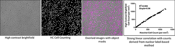
Image: The high contrast brightfield (HCBF) method delivers accurate label-free cell counts comparable to methods relying on nuclear dyes, without disrupting cells. Image courtesy of Biotek.
Although HCBF is typically used for cell counting applications, “we can use fluorescent probes common to phenotypic assays (reactive oxygen species, mitochondrial membrane potential, apoptosis, etc.) in conjunction with HCBF to normalize data to the total number of cells in the assay well. This tends to improve precision in the experimental data,” says Banks.
He highlights that BioTek’s techniques can be used beyond just cell proliferation, since in general, labels can disrupt cell behavior. “BioTek developed for use a number of label-free techniques applied to cells in many more applications than cell proliferation. These include brightfield, HCBF cell counting, and Zernike-based phase contrast.”
And Dodla adds that label-free proliferation assays are becoming more widespread. “Advances in imaging technologies are making this technique more accessible to labs around the world.” However, he cautions that these assays may require the purchase of additional instruments and software analysis tools for high contrast counting.
Cell proliferation experiments are usually time consuming, and cells must remain healthy throughout the duration. “Typically at the beginning of the experiment perhaps a few thousand cells are seeded into each well of a 96-well microplate. It takes about 2–4 days, depending on the cell line and experimental conditions for the wells to be confluent. It is imperative that the cells remain healthy throughout this duration and this is best performed by providing the environment control of a CO2 incubator,” says Banks. He notes that instead of manually collecting a microplate from an incubator and imaging it periodically, some researchers might prefer an instrument like BioTek’s BioSpa 8. “This automated CO2 incubator shuttles up to 8 microplates sequentially to our environment controlled imagers. This allows full walkaway automation through the duration of the experiment.”
And it’s user friendly. A simple optimization step defines the correct focal heights for acquiring HCBF images, says Banks. Once there is a defined protocol, the system will take it from there, automatically taking images and performing quantitative analysis over the course of the experiment. He says that these protocols can then be saved for future use with the same or similar cells. “By providing both percent confluence and cell counts, researchers are able to more fully evaluate treatment effects on proliferation rates and changes to cell morphology within a single assay.” BioTek also offers the Lionheart automated imager that has up to 100X magnification and supports a range of assays, including cell proliferation.
Less toxic and more sensitive reagents
If you’re not in the market for imagers or fully automated systems, Dodla says there other nontoxic proliferation assay options, as well. “Proliferation assays such as those based on variations of MTT, allow for continuous monitoring of cells without cytotoxic effects.” He’s referring to the nonradioactive colorimetic assay based on WST-1, which comes by itself or in a convenient CCK8 kit as a ready-to-use solution that also contains an electron-coupling reagent. WST-8 is reduced by dehydrogenase enzymes in cells and is more stable and less toxic than the other tetrazolium salts. According to the MilliporeSigma site, because of this lack of cytotoxicity it can be used for longer incubations of up to 48 hours.
MilliporeSigma also offers several options for live cell dyes for long-term use, including SE and CFSE BioTracker kits, says Dodla. These initially non-fluorescent esters diffuse into cells and are converted to fluorescent dyes by intracellular enzyme cleavage. These dyes can be used to track cell divisions either in vitro or in vivo by monitoring the fluorescent intensity that decreases with each cell cleavage.
In addition to the high contrast imaging described in this article, there are other types of label-free proliferation assays available “such as those based on monitoring electrical impedance to predict cell number, adhesion, viability, and morphology changes,” says Dodla. And he points out that there are now 3D cell proliferation assays that allow for cell counting in 3D spheroids or matrices. Banks notes that HCBF works for spheroid proliferation applications using ULA microplates.
But in the end, Dodla says that you will need to optimize whatever proliferation assay you choose to work with your cell type. “Also, most proliferation assays do not identify the mechanisms behind changes in proliferation. Additional assays might be needed to understand the mechanisms for change in proliferation rates.”