Immunophenotyping has traditionally been synonymous with immune cell characterization by flow cytometry. This technique of using fluorescently labeled antibodies to detect and quantify defined cell populations within a suspension sample has relied on established methods for many years. A drawback of these approaches is that they are often susceptible to inter-assay and site-to-site variability, leading to uncertainty over results. Moreover, conventional methods frequently have high sample volume requirements to deliver only a limited number of readouts without spatial context. To address these issues, immunophenotyping technologies have had to evolve.
A new approach to cell enrichment improves yield
Variability can be introduced at multiple stages within an immunophenotyping experiment, not least during the initial isolation and concentration of white blood cells (WBC) from whole blood. This is typically performed by density gradient centrifugation, a labor-intensive process that can artificially skew results. “During density gradient centrifugation, cells experience significant centrifugal forces with concurrent exposure to chemical reagents,” notes Anya Manning, senior product manager at MicroMedicine. “This can influence cellular quality or result in the loss of cells before you even begin your assay. Density gradient centrifugation also narrows potential downstream analysis by eliminating polymorphonuclear cells such as granulocytes, retaining only monocytes and lymphocytes for subsequent immunostaining.”
To overcome these problems and reduce contaminating events such as red blood cells (RBC) and platelets, MicroMedicine has developed an automated, novel technology for enrichment of all nucleated cells from whole blood. Based on inertial focusing microfluidics, the Sorterra™ system automates cell separation and concentration to improve yield and purity of viable WBCs over Ficoll®-based methods. “Our technology centers around a microfluidic disc that leverages small differences in cell size to quickly and gently separate cells with less than five minutes of hands-on time,” explains Manning. “We recently presented a poster at SITC showing 25% more viable PBMCs recovered and 10x greater elimination of contaminating RBCs and platelets compared to density gradient centrifugation. Furthermore, ELISpot B and ELISpot T data showed equivalent functional responses for both isolation methods.”
Metal-labeled antibodies simplify panel design
Continued evolution of immunophenotyping platforms that enable higher parameter analysis combined with robust reproducibility has culminated in mass cytometry—a variation of flow cytometry that relies on antibodies labeled with metal tags to deliver high-multiplex immune profiling capabilities. “Mass cytometry has seen increased uptake for immune profiling as awareness of the benefits of metal-labeled antibodies continues to grow,” reports Michelle Poulin Ph.D., senior manager, mass cytometry applications North America at Fluidigm. “Metal-labeled antibodies are very stable, and the tags are not affected by different fixation or permeabilization methods the way fluorochromes can be. There is also little to no signal overlap, meaning signal compensation is not required. Furthermore, because the metals Fluidigm uses as tags are not naturally occurring in biological systems, there is no interference from native background or autofluorescence. In combination, these features greatly simplify panel design.”
Recent advances in mass cytometry are epitomized by Fluidigm’s 2017 introduction of the Hyperion™ Imaging System, a platform that has enabled characterization of the tissue microenvironment at subcellular resolution using Imaging Mass Cytometry™ (IMC™). Poulin notes that, today, IMC is a well-published high-multiplex application for phenotyping and functional analysis of complete cellular systems in tissues and tumors using familiar IHC protocols. Fluidigm has supported this growth by developing a range of Maxpar® Human Immuno-Oncology IMC™ Panel Kits; these provide a modular strategy for researchers to build upon and customize, allowing a quick start and reducing time and cost to ramp up research.
A one-tube mass cytometry assay minimizes variability
According to Poulin, additional factors driving adoption of mass cytometry are greater accessibility and better standardization. She cites as an example the company’s Maxpar Direct™ Immune Profiling Assay™, a dry 30-marker antibody panel provided in a single tube, which improves immunophenotyping standardization in several ways. “First, the antibody panel is based on the recommendations of the Human Immunology Project Consortium (HIPC), a centralized knowledge base dedicated to immune profiling,” she says. “Second, since sample (human whole blood or PBMC) is added directly to the tube, any pipetting differences between technicians are removed. Another advantage of this assay, and mass cytometry in general, is that no assay-specific optimization is required. Once the staining protocol is complete, samples are acquired on the Helios™ mass cytometer using an assay-specific acquisition template. Because any Helios mass cytometer undergoes the same daily QC and an internal beads standard is added to each sample, no optimization or standardization is needed before samples are run on multiple instruments or across multiple sites.”
Tissue-based immunophenotyping provides spatial context
These days, immunophenotyping is no longer limited to suspension samples. Instead, novel approaches based on familiar IHC workflows provide spatial context in terms of tissue architecture. “By performing immunophenotyping in situ, researchers can truly determine whether cell-cell interactions are taking place,” notes Dr. Sean Downing, director of applications, pathology and services at Ultivue. “Multiplex IHC has moved the mindset of a one biomarker-one phenotype paradigm to a mode of tissue-based immunophenotyping similar to that currently performed by flow cytometry. Now, it is possible to identify multiple cell types in tissue simultaneously, measuring not only their abundance but also their spatial activity in the context of the microenvironment. This could provide novel insight not previously obtainable from a cell suspension.”
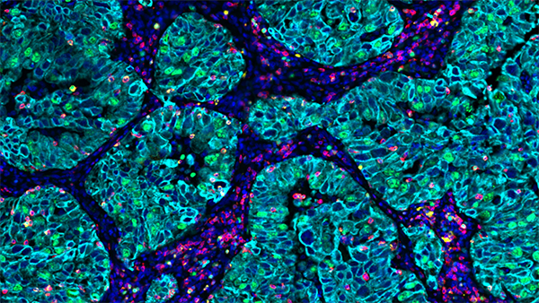
Image: UltiMapper T cell activation panel staining NSCLC tissue – CD3 (red), Granzyme B (green), Ki67 (orange), CK (cyan). Image courtesy of Ultivue.
The value of tissue-oriented immunophenotyping is highlighted in a recent publication by Rimm et al, where the authors used Ultivue’s InSituPlex® DNA-barcoding technology to study the tumor microenvironment and correlate PD-L1 expression in macrophages with overall survival in NSCLC patients. An advantage of this method is its compatibility with instrumentation and software found in most IHC laboratories, allowing for integration into virtually any histology workflow. “Using DNA-barcoded antibodies improves immunophenotyping standardization by eliminating the need for secondary antibodies,” reports Downing. “Moreover, our approach maintains biomarker ratios by linearly amplifying all the DNA barcodes at the same rate and at the same time; this provides more accurate downstream image analysis than geometric amplification methods that are temporally separated.”
Industry-wide efforts toward immunophenotyping standardization
Complementing the HIPC program, the 10,000 Immunomes Project created at the University of California, San Francisco collects and annotates data from multiple platforms to provide a reference dataset for human immunology. This includes flow cytometry, mass cytometry, multiplex ELISA, gene expression data, and more, taken from more than 10,000 healthy subjects. “These collective resources provide researchers with an extensive baseline reference of the diversity in healthy human immunology and can be used in future studies to compare normal and disease states,” explains Clinton Hupple, product manager, proteomics at Fluidigm. As industry-wide efforts such as these continue to evolve alongside various enabling technologies, researchers will benefit from further improvements to immunophenotyping standardization.