Exosomes are tiny, but their potential is mighty. The smallest of the small extracellular vesicles (EVs), these lilliput travelers carry cargo (i.e., cellular information) in both their plasma membranes and cytoplasmic cores. They can be thought of as a type of descendent of cells, steadily budding off, infused with biomolecules that offer a readout of the activity occurring within the original cell.
“There is still a lot we don’t know about exosomes and other small EVs, but we do know that exosomes are released under normal physiological conditions and play an important role in intercellular communication by transferring cargo between cells,” says Haley Pugsley, senior scientist at Luminex Corporation. “This cargo may include proteins, DNA, and RNA, including miRNAs. The release of exosomes is also a way to get rid of unwanted molecular material in the cell.”
An increase in the rate of exosomal release or altered expression of the cargo can be a warning sign that something has shifted or that disease is present. “When exosomes are released by cancer cells they can act as circulating biomarkers for that cancer,” says Pugsley. Scientists have already started to find patterns in the types of molecules harbored by exosomes and specific cancers.
“Guo et al. showed [exosomes] are involved in tumorigenesis, metastasis, and drug resistance. We suspect that they are information carriers communicating with other tumor cells in the microenvironment,” explains Yasha Talaga, applications & collaborations product manager in cell biology for Bio-Rad.
Recent work finds that exosomes work in tandem with the primary tumor to condition the microenvironment and promote growth both near and far. For example, one group found that in chronic myeloid leukemia, tumor-derived exosomes (TDEs) rich in cytokines could be transported to nearby tumor cells, activate proliferation and enhance cancer cell survival mechanisms. Similar mechanisms were described in other types of cancers. Gastric cancer TDEs use miR-27 to induce proliferation in adjacent cells, and prostate cancer TDEs rely on long non-coding RNA c-MYC-Upregulated to instigate tumor growth. And the list continues to grow.
As a system of transport, intercellular communication, and waste renewal, exosomes are shed into nearly every type of bodily fluid, including blood, urine, and saliva. Given the ubiquity of exosomes, and easy accessibility to them, it’s easy to see why this research is getting so much attention and why there is hope that these novel biomarkers will be powerful, noninvasive diagnostic, monitoring, and even treatment tools. But first scientists need to figure out the best way to isolate and characterize these nanoscale transmitters.
Separating out exosomes from the noise
The small size of EVs, which include apoptotic bodies, microvesicles, and exosomes (in the nanometer range), makes studying and characterizing them challenging. Isolating them involves a tedious process that relies on a multitude of techniques, including ultracentrifugation, density gradients, size exclusion, and precipitation. Flow cytometry seems to be the method of choice for most scientists, says Pugsley. “ [It] is a great tool for this as it can immunophenotype a complex mixtures of EVs.” Although an enrichment step such as ultracentrifugation is still usually recommended prior to sorting.
Bio-Rad’s ZE5 cell sorter can isolate exosomes without the ultracentrifugation step, according to Talaga. “The ZE5 makes it easy to resolve these small particles from electronic noise or sheath [the solution that runs through the flow cytometer] impurities.” The analyzer is equipped with a small particle detector (with FSC [Forware SCatter] PMT from the 405nm laser). Talaga notes that the ZE5 can detect exosomes without beads, although Bio-Rad does offer bead conjugation methods. Bio-Rad carries validated exosomal markers such as CD63 and CD81, as well as intravesicular markers like ALIX.
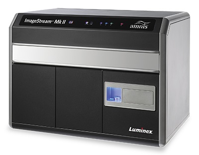 Luminex also has flow cytometry options for small particle detection. The ImageStream® X Mk II and the CellStream® flow cytometers can detect fluorescent EVs. The former can also monitor EV binding and internalization in cells. With fluorescently labeled EVs, Pugsley says characterization in biofluids is feasible and researchers can determine where they originate from (specific tumors, for example). They can also determine whether they bind to or are internalized by other cells.

Pugsley advises being diligent with controls. “EVs are very small and at the limit of detection for many techniques, including flow cytometry. [Controls should include] antibody/dye only, buffer only, EV only (no fluorescent label), and labeled EVs plus a detergent, like Triton X-100. Running dilution series of your labeled EV samples as well as the controls will help ensure that you are in fact measuring EVs and not artifacts.”
And Talaga points out that sheath solutions can carry small particles, which can confound exosome research. “Steve McClellan, at the University of South Alabama, has mentioned that he has graduate students who spend six hours filtering their sheath fluid before it is put on the flow cytometer! In our system, that becomes unnecessary because we have on-board sheath filters.” It’s worth noting that the ZE5 uses deionized water as its sheath. Sheath purification, filtered buffers, and ensuring proper set-up of the flow cytometer are critical for successful exosome detection, says Talaga.
Exosome Isolation and Analysis kits from Abcam are another option. “They enable straightforward and optimized exosome separation from cell culture and fluids such as plasma, which is critical for the development of exosome analysis in clinical research,” says Michael Tackett, assay development director for Abcam. These are bead-based assays that use pre-enriched ultracentrifuged exosomal populations. “These kits are handy for examining exosome populations using flow cytometry for consistent, reliable results.”

Image: NIH/3T3 (mouse embryo fibroblast) cells showing vesicular cytoplasmic localization of exosome marker CD63 following staining with anti-CD63 antibody (ab217345) and Alexa Fluor®488-conjugated secondary (ab150077, green). An anti-alpha tubulin antibody [DM1A] conjugated to Alexa Fluor® 594 was also used (ab195889, red), with DAPI nuclear counterstain (blue). Image courtesy of Abcam.
miRNA biomarker signatures
All cells, both healthy and diseased, shed vesicles (including exosomes) into bodily fluids, which makes them appealing for use as diagnostic indicators. In recent years, they’ve emerged as biomarkers for cancer detection and progression. The architecture of the exosome protects RNA and miRNA from degradation by RNase catalytic activity. As such, exosomes function as an intact snapshot or descendent infused with the traits of the original tissue or tumor. MicroRNAs and, in particular, distinct miRNA signatures, have been identified and attributed to specific cancers. However, the multiplexing technology required for garnering such signatures is typically stymied by sample availability. And miRNA purification can alter the miRNA profile, yield, and throughput.
Abcam’s FirePlex® particle technology, which can detect miRNA directly from crude sample digests, is an appealing option, cutting out cumbersome steps that can introduce user error. According to Tackett, up to 70 analytes per well can be detected with the FirePlex® miRNA assays. “These provide multiplexed miRNA measurements from only microliters of biofluid including blood, serum, plasma, spinal fluid, and urine, and the exosomal fractions of these fluids (irrespective of enrichment methodology), all without the need for RNA extraction” These assays can be carried out on standard flow cytometers and yield results comparable to qPCR- and microarray-based approaches.
Pugsley is most excited by the prospect of EVs and exosomes being used as cancer biomarkers for early disease detection, but she says we still have challenges to overcome. “Some of the major difficulties facing the EV community include the lack of standardization of protocols for EV enrichment and characterization making it difficult to interpret the data from different studies.”
Exosome Isolation Techniques
- Ultracentrifugation (spinning samples at forces up to 1,000,000G) is thought to be the gold standard for exosomal isolation. Relies on differences in density and size. Can yield large amounts of exosomes when done correctly (pay attention to washing steps and be sure to perform at 4°C to keep proteases, DNases, and RNases inactive). Often used in combination with sucrose gradients. Very labor intensive.
- Precipitation-based methods utilizes solubility differences to extract exosomes. Kits available. Can be used with small volumes on variety of bodily fluids. However, may not be good for downstream mass spectrometry work.
- Size exclusion-based methods, such as ultrafiltration and size exclusion chromatography, use differences in size between exosomes and other particulates. Kits available. Relatively fast and easy, however, may only offer moderate exosome purity.
- Immunoaffinity-based techniques may rely on antibodies conjugated to beads. Validated antibodies and kits available. Can isolate specific exosomes. Works well with flow cytometry. Can be an expensive method.
- Microfluidics-based isolation techniques can use physical and biochemical properties of exosomes in combination with acoustic, electrophoretic, and electromagnetic manipulations. Can be used with very small sample volumes and limits reagent consumption. However, suffers from lack of standardization, method validation.