Super resolution microscopy (SRM) takes the resolution of optical microscopy below the diffraction limit of light. This allows visualization of cellular details that could not previously be seen without using alternative techniques such as electron microscopy. Despite increased uptake in research areas including cell biology, neurobiology, microbiology, and drug discovery, the use of SRM has been limited by high ownership costs and a lack of well-established protocols and analysis methods. Now, with a variety of affordable, user-friendly SRM platforms entering the market, accompanied by a growing list of publications validating their utility, SRM looks set to become mainstream.
Changing research needs are driving development
According to Leanna Ferrand, global product support leader at GE Healthcare, two key research demands are driving developments in SRM: the need for longer and faster live-cell imaging and the desire to image thicker, more complex samples.

“Scientists are increasingly employing live-cell samples to capture dynamic biological processes in real-time,” she says. “There is also a growing trend to study composite sample types such as organoids, mixed cell cultures, and tissue samples. To support this research, SRM techniques are evolving to span the total duration of cellular processes and stand up to the challenges of imaging deeper into any sample without compromising biological relevance. In the past, researchers have had to accept partial answers when the diffraction limit of traditional microscopy tools did not provide the level of detail required for complete understanding. Now, they are no longer limited by the tools available, they just need to ask the right questions.”
Image: Visualizing HeLa cell division. Localization of Kif18B (purple) and EB1 (orange) at the very end of a 25nm diameter microtubule (blue), a component of the mitotic spindle. DNA is pictured in green. Image courtesy of GE Healthcare.
SRM is seeing widespread adoption
“Since the advent of SRM, a lot of work has gone into making the techniques more accessible to the scientific community,” notes Renée Dalrymple, sales development manager, 3D light microscopy solutions at ZEISS Research Microscopy Solutions. “For example, the ZEISS Airyscan changed confocal microscopy by allowing researchers to not only accomplish super resolution—down to 120 nm in the lateral (xy) plane and 350 nm in the axial (z) plane—but also to image more gently than with traditional confocal microscopy by using more of the fluorescent signal from the sample. More recently, we’ve added the Elyra 7 with Lattice Structured Illumination Microscopy (SIM) to our portfolio. The lattice pattern provides a dramatic improvement over traditional SIM, which uses a stripe pattern. In providing these platforms, Zeiss is contributing to wider adoption of SRM across many different research fields. This is exemplified by the fact that, a few years ago, researchers began telling us that their papers were being rejected due to reviewers asking why they hadn’t tried their experiments using SRM.”
 “SRM has really gained in popularity as the technologies have transitioned from DIY environments to commercially produced microscope systems,” adds Lauren Alvarenga, product manager for life science microscopy at Olympus Corporation of the Americas. “Today, super resolution microscopes like the Olympus SpinSR10 can be run in exactly the same way as a simple, widefield microscope, eliminating some of the barriers presented by complicated imaging techniques. A further advantage of the SpinSR10 model is that it allows switching between widefield, confocal, and SRM imaging modes with no need to change samples, imaging buffers, or fluorophores. This makes it ideal for multi-use environments like core imaging facilities, where SRM can complement other imaging modalities.”
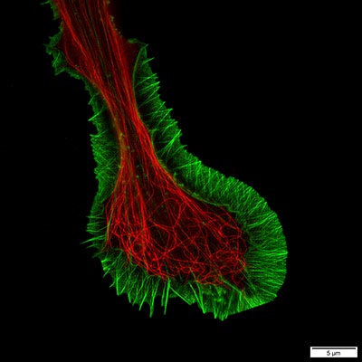
Image: Fluorescent staining of microtubules (red) and actin (green) in growth cone of NG108 cells, imaged using Olympus IXplore SpinSR. Image courtesy of Dr. Kaoru Katoh, Biomedical Research Institute, National Institute of Advanced Industrial Sciences and Technology (AIST).
Key considerations when choosing an SRM technique
- How much spatial resolution is needed?
- How much temporal resolution is needed?
- Are the samples live or fixed?
- How deep into the sample do you need to image?
- How many channels do you need to image?
- What field of view is required?
- What sample preparation tools do you have available?
Providing quantification at the single molecule level
Alexander Carr, consultant at TTP plc, reports that in addition to allowing smaller structures to be visualized, a major advantage of SRM is its compatibility with quantification, especially at the single-molecule level. To elaborate on this point he explains that, broadly speaking, there are two categories of SRM techniques, each of which fulfils a different purpose. “Deterministic SRM techniques such as STED and SIM exploit nonlinear responses (e.g. stimulated emission or saturation of excited states) to gain additional spatial information,” he says. “Stochastic SRM techniques like PALM and STORM instead use the ability of some fluorophores to switch between a ‘bright’ and a ‘dark’ state to locate the position of each fluorophore in the sample with nanometer-scale precision. While deterministic SRM typically offers faster imaging speeds, allowing real-time videos to be recorded, the power of stochastic microscopy (also known as single-molecule localization microscopy, or SMLM) is in the nature of the information recorded. Rather than producing an image, SMLM generates a list of coordinates detailing the positions of individual fluorophores within the sample. This is readily compatible with quantification of metrics, making SMLM ideal to measure complicated diffusion dynamics, characterize clustering, or determine co-localization of different targets at a biologically relevant scale.”
Multiplexing through new dimensions
“A current trend in biomedical research is for more markers to be visualized simultaneously, especially in living samples, and SRM is certainly delivering on this need,” notes Ryan Hrejsa, senior marketing manager, life science research at Leica Microsystems. “For example, our SP8 scanning microscopes can register up to five spectral SRM channels simultaneously and, used in combination with fast lifetime contrast (FALCON) technology, can incorporate an additional dimension of fluorescent lifetime imaging (FLIM) into protocols. This allows researchers to perform biosensing and to accurately track multiple fast molecular interactions.” The value of FLIM is illustrated in a publication by Lanzanò et al, in which it significantly improved the resolution in STED analysis of live cells.
The Lanzanò publication is just one of a growing number of articles highlighting the importance of SRM to support a diversity of research. Other examples include its use to examine the organization and dynamics of plasma membrane components; its application in studying the function of the cell nucleus; and its utility to help provide structural understanding of a protein in the sub-20 nm range. SRM has also been fundamental in identifying the essential role of a protein complex during Plasmodium falciparum invasion of erythrocytes in malaria, and has proven utility in studying neuron plasticity.
Entering mainstream research
“The research areas where we see the highest uptake of SRM techniques all have one thing in common: a scientist who was brave enough to incorporate SRM into their research,” says Ferrand. “These early adopters have been pivotal in demonstrating the power of SRM in their field through publications and conference presentations; in asking just a single question of SRM, they have uncovered hundreds of new questions they never knew to ask before.”
Carr adds that as adoption increases, more trust will be placed in SRM results, creating greater incentive to provide standardized protocols and novel reagents. “I expect that the ability of SRM to analyze heterogeneity within samples will find many applications in diagnostics and big pharma,” he says. “This is especially true for cases in which subtle changes of small sub-populations within a sample can lead to significant effects. Examples of these cases include protein aggregation in neurodegenerative diseases such as Parkinson’s and Alzheimer’s, as well as drug binding and protein binding kinetic studies. I am excited to see new techniques emerge and be involved in all the amazing applications that will be found as SRM enters the mainstream.”