In microscopy, scientists select from a collection of options. One of the most common choices is: widefield or confocal? Despite this being such a common question, some experts say that scientists could often make a better choice. It all depends on the application and the required data, as well as data quality.
“In general, a widefield microscope is easier to use, less complicated and cheaper than a confocal microscope,” says Nathaniel Peters, administrator of the University of Washington Keck Microscope Facility. “The optical components of the microscope stand itself are the same for widefield and confocal, but the real difference lies in the nature of the light used for excitation, how the sample is illuminated, and how the image is collected.”
In a widefield microscope, the entire focal volume is illuminated, but that creates blur from areas out of focus above and below the image plane; a confocal microscope scans a sample with a focused beam of light, more than one beam in some platforms. Both methods provide benefits to scientists imaging samples, but one or the other is a better choice in most situations.
“Straight vanilla widefield microscopy has its place in science and research, but the real benefit and significant improvement of widefield comes from combining an automated widefield microscope with restorative deconvolution algorithms to enable imaging of biological samples that fall into the categories of small, dim, and/or alive,” says Leanna Ferrand—global product support leader, genomics and cellular research, GE Healthcare Life Sciences. So, this method works for various samples—alive or fixed—and even in dim light. Widefield microscopy also works well in dynamic situations, such as imaging moving specimens. The disadvantages of widefield microscopy are “that it does not do well with thick specimen or samples that scatter light significantly,” Ferrand notes.
For thicker samples or ones that really scatter light, Ferrand recommends confocal microscopy. “The background rejection from confocal techniques is helpful for tissue, thick specimens, or even in a high-content approach,” she explains. “This saves you time as you can, for example, skip final washing steps, because you’ll reject the background noise and so on.”
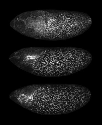
Image: This composite image shows a late-stage developing Drosophila egg chamber from a fruit fly ovary expressing E-Cadherin::GFP, which is a cell-cell adhesion molecule. Over about 6 hours, a z-series (about 50-micron sections) was acquired on a laser scanning confocal microscope, and these images are maximum intensity projections of those. Image courtesy of Nathaniel Peters from research done in the laboratory of Celeste Berg at the University of Washington.
The main difference in resolution between these types of microscopy comes, not in the x-y plane, but along the z-axis. In fact, closing down the pinhole used in confocal increases the resolution in depth, but that sacrifices the level of light used to form the image. “Olympus confocals are so sensitive that you can close down the pinhole and still get a good image,” says James Lopez—manager, life science applications group at the Olympus Corporation of the Americas. For something moving, though, he says scientists will often get a better image with “widefield because of the larger optical-section thickness.”
Five Fast Facts about Widefield Microscopes
1. A widefield microscope is easier to use, less complicated, and cheaper than a confocal microscope.
2. Widefield microscopes enable imaging of biological samples that are small, dim, and/or alive.
3. Widefield microscopy works well in dynamic situations, such as imaging moving specimens.
4. A disadvantage of widefield microscopes is that they do not do well with thick specimen or samples that scatter light significantly.
5. Widefield systems are easier to house and maintain, and fine as long as the sample is thin—for example, a single layer or cultured cells.
If a scientist wants a rule of thumb for making a choice between these kinds of microscopy, Lopez says, “A thin sample usually means widefield; tissues usually mean confocal.” Still, even if people know that simple concept, it’s not always enough. “People stay with what they know and often use the wrong technique,” Lopez says. “Once people own the equipment, they often use what they have and do not consider alternatives.”
Just because a piece of equipment is already in a lab doesn’t make it the best choice for every imaging situation.
Available options
The size of the microscope market means that one article can only consider a few options among the existing platforms. Let’s look at a couple products mentioned by the experts interviewed here.
For recent advances in widefield microscopy, Ferrand points out GE’s DV Ultra. This scope “delivers scalability to a widefield microscope,” she says, “enabling the same powerful widefield imaging with deconvolution for single slides or samples with the ability to scale up to multi-well plate formats.”
Five Fast Facts about Confocal Microscopes
1. Confocal microscopy is recommended for thicker samples or ones that really scatter light.
2. The background rejection from confocal techniques is helpful for tissue, thick specimens, or even in a high-content approach.
3. Some confocal platforms can be used effectively on live specimens as well as live-cell imaging.
4. Confocal is the clear best choice in some cases, like with tissue slices.
5. Confocal microscopy provides more resolution in the z-axis, or depth.
For advances in confocal microscopes, Ferrand mentions GE Healthcare’s EDGE Confocal on the IN Cell 6500. As she explains, this “builds on to our IRIS Line scan confocal technology providing point-scan confocal image quality at spinning-disc confocal acquisition speeds.” She adds, “EDGE improves enhanced background rejection and finer z-sectioning optically.”
Some of the advances will surely drive new capabilities. Sometimes, even the experts wonder about the possibilities. “What kinds of scientific discoveries could be enabled,” Ferrand asks, “if we combined the power of EDGE imaging with super-resolution techniques that GE Healthcare is already enabling scientists to utilize on the DeltaVision OMX SR?”
Some past constraints are already disappearing. For instance, scientists can use some confocal platforms effectively on live specimens. For instance, the Olympus FLUOVIEW FV3000 series is fast enough for live-cell imaging. So is the Airyscan, says Scott Olenych—North American product marketing group manager, light microscopy at Carl Zeiss Microscopy. “It has a special detector that is arranged for better resolution and a much higher signal-to-noise ratio,” he explains.
Souped-up software makes a big difference in today’s imaging, as well. For example, the Olympus cellSens Dimension deconvolution software works with graphic processing units (GPUs). “It can do deconvolution on widefield, confocal, two-photon, and spinning-disk microscopy,” Lopez says. “Historically, deconvolution was not considered necessary with confocal, but it’s worthwhile with the improvement in the images.”
Automating imaging
Like most of science, today’s imaging is often driven to handle more information, and microscope companies need to fuel the options. “We’re moving toward large volumes of data and better analysis,” says Olenych.
As an example, Olenych points to the Celldiscoverer 7, which is an automated platform for live-cell imaging. “It’s an automated, inverted format,” Olenych says. “You just load the samples in 96-well plates, individual dishes, slides—any live format.”
For automated slide scanning, scientists can use the Zeiss Axio Scan.Z1. “A robot loads the slides, there’s some autofocusing, then the image is scanned at an appropriate magnification and the image is archived,” Olenych explains. “It can handle up to 100 slides.”
Brief review
It’s worth remembering—and being reminded of—the basics in this common microscopy decision. “Widefield systems are more affordable, great for speed, easier to house and maintain, and fine as long as the sample is thin—for example, a single layer or cultured cells,” Peters says. “Confocal microscopes are very expensive and large, require more significant training to operate, and are generally slower to use.”
Still, confocal is the clear best choice in some cases, like with tissue slices. Here, says Peters, “the ability to exclude out-of-focus light and take serial sections through a sample provides a tremendous advantage in z resolution, or depth.”
The key is matching the best method to a particular problem. Instead of just using what’s in a lab, it’s worth considering what would work the best, and then getting access to that kind of microscopy and learning to use it.
Image at top: Confocal imaging can be used to make a series of images going down through a sample. GE Healthcare’s EDGE Confocal allows very fine vertical optical sections. Image courtesy of GE Healthcare Life Sciences.