Flow cytometry assays have traditionally been used to determine the percentage of various cell types within a population; to assess cell health functions such as apoptosis, viability, cell cycle, and proliferation; and to study protein expression and activation status. Yet the wealth of information that can be gained from flow cytometry has grown significantly in recent years following demands to increase the number of simultaneous measurements, elevate throughput, and deliver more complex readouts.
One company helping researchers to maximize the potential of their flow cytometry assays is BioLegend, which offers a wide selection of antibody conjugates. Supporting these with open-access online resources, including a spectra viewer and multicolor panel builders, BioLegend recognizes the need for fluorescent reagents to keep pace with advances in instrumentation. “We’ve watched multicolor panels grow to upwards of 20+ parameters,” says Ken Lau, technical marketing manager. “Several years ago, our introduction of Brilliant Violet™ fluorophores greatly enhanced the utility of the violet laser, expanding options in a multicolor panel. As cytometers become more sophisticated, the demand for fluorophores that can fill these new channels increases, and we’re continuing to work hard to meet these needs.”
As cytometers become ubiquitous, they’re being used in novel applications, even building on the typical ELISA immunoassay. While most ELISAs require one analyte to be analyzed per plate, BioLegend’s LEGENDplex™ technology provides a more robust solution. This bead-based immunoassay is compatible with standard flow cytometers and run in microplates, allowing for the analysis of 13 targets simultaneously. “When you perform ELISAs, you inevitably hit a cap with how many plates you can juggle at once. LEGENDplex cuts down on time, reagents, and cost, which is obviously helpful when research deadlines are fast-approaching,” says Lau. “Flow cytometry is a versatile tool, so we’re adapting it to many platforms like tetramers, screening, and cell sorting. It’s an exciting time as we keep pace with advancing research.”
Scaling up
Also addressing the need for scale-up by enabling plate-based flow cytometry, Thermo Fisher Scientific has introduced instruments such as the Attune™ NxT flow cytometer and the Attune NxT autosampler to their portfolio. “Plate-based instruments such as the Attune NxT flow cytometer significantly reduce the amount of sample material and reagents required, while automation provides the added benefit of freeing up time following set up,” explains Dr. Helen Fleisig, product manager, flow cytometry reagents.
Biocompare’s Flow Cytometer Search Tool
Find, compare and review flow
cytometers from different suppliers Search
There is undisputed value in studying protein expression patterns of cells, however, not placing equal importance on the underlying dynamics of RNA transcription and the impact it can have on both protein expression and cell function leaves us with an incomplete, one-dimensional view. “A major advantage of our PrimeFlow™ RNA assay, launched recently, is that it adds a second dimension to cell studies,” says Fleisig. “PrimeFlow detects mRNAs and represents the first flow cytometry assay capable of simultaneous RNA and protein detection.”
Combining the power of branched DNA technology with the single-cell resolution of flow cytometry, PrimeFlow enables detection of up to four RNA targets in combination with immunophenotyping for cell surface and intracellular proteins using fluorochrome-conjugated antibodies. “While PCR and sequencing can provide comprehensive gene expression data in bulk sample preparations, the analysis of bulk samples can mask the individual effects of unique cellular subsets,” explains Fleisig. “Using our PrimeFlow RNA assay to amplify specific RNA transcripts, researchers can analyze millions of cells from specific sub-populations for unique transcript expression levels. It is also possible using PrimeFlow to evaluate transcriptional regulation and protein expression over time.”
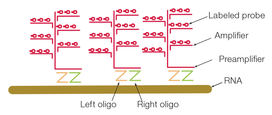 Image: The PrimeFlow RNA Assay expands the capability of flow cytometry to measure RNA. A gene-specific oligonucleotide target probe set binds the target RNA sequence prior to amplification. Detection utilizes fluorescently labeled probe oligonucleotides. Image courtesy of Thermo Fisher Scientific.
Higher complexity
Similarly keen to stress the importance of generating data from individual cells rather than an overall suspension of cells is Maria C. Gentile, global marketing manager at Beckman Coulter Life Sciences. “Flow cytometry is unique in the way that it can process multiple parameters per cell while maintaining the cellular context of each measurement,” she says. “Techniques such as Western blotting and ELISA provide only average responses, showing cellular correspondence but lacking phenotype, while microscopy is much slower than flow cytometry at processing comparable cell numbers. Beckman Coulter have been committed for decades to the drive toward higher complexity in flow.”
CytoFLEX technology and DuraClone kits are just two examples of Beckman Coulter’s many innovative flow cytometry products. “Our CytoFLEX flow cytometry platforms employ a revolutionary detector technology to host up to 23 parameters from 6 lasers within an exceptionally small footprint,” explains Gentile. “By using side scatter from the violet laser line, these instruments provide advanced sensitivity and superior resolution for small particles, affording particle detection to 80 nm relative to polystyrene particles.” Ideally suited for use on the CytoFLEX flow cytometers, DuraClone kits are provided as dry antibody panels of up to 10 colors, pre-formulated in single tubes. These are easy to implement, room temperature-stable, and cover immunophenotyping, immune functional assays, and the detection of rare malignant leukocytes. “The recent addition to the DuraClone family, three DuraClone RE antibody panels optimized for the detection of abnormal rare CD5+ B cells, plasma cells, or immature B cells, were launched last year,” adds Gentile.
Fluidics-free systems
Fluidics-free systems not only reduce the amount of sample material that is required, but also allow for imaging of infectious material without the need for instrument decontamination, as well as permitting accurate analysis of clumpy or very fragile cells. Timothy Smith, product manager at Nexcelom Bioscience, describes how these systems, based on image cytometry, are a popular alternative to flow cytometry. “Image cytometry performs experiments similarly to flow, but without the issue of clogging and with no need for cleaning, calibration, or maintenance,” he says. “By taking an image of the cells, then using image processing algorithms to determine cell number, size, shape, and fluorescent emission, a similar range of assays to those for which one would use a flow cytometer can be performed.”
Nexcelom Bioscience’ Celigo, a benchtop microplate-based brightfield and fluorescent imaging instrument, provides high-speed, fully automated imaging of suspension or adherent cells, tumor spheroids, iPSC, and cancer stem cell colonies in plates, flasks, or slides. The company has also recently added the Spectrum, a versatile cell-counting and cell-based assay system that utilizes fluorescent reagents and kits, to their Cellometer product range. “Both of these instruments provide visual verification within images that every cell in a well has been identified and counted,” adds Smith. “Results and analytics, gating, and multi-color scatter plots and histograms are comparable to those of flow cytometers. In general, any assay that is run on a flow system can be adopted to run on an image-based instrument.”
“In my opinion, the next big wave of innovation will start with the cytometers,” notes Lau. “Several companies are now creating spectral cytometers, capable of distinguishing fluorophores with similar emission spectra to detect over 48 parameters.” According to Gentile, advancement of technologies for nanoscale flow cytometry is also progressing, driven by research in the areas of exosomes and extracellular vesicles. Fleisig suggests that flow cytometry assays targeting metabolic functions will help provide detailed answers to complex questions. Whatever the direction, new product development will continue to support the flow cytometry assays of the future.”