Advances in microscopy cameras over the past decade may now mean that for the vast majority of bioscience applications, laboratories can get away with investing in just a single high-performing camera, says one of the field’s leading experts, Simon Watkins, Distinguished Professor and Vice Chair of Cell Biology, Professor Immunology, and Founder and Director of the Center for Biologic Imaging at the University of Pittsburgh.
“The best solution for nearly all bioscience uses today is the monochrome scientific CMOS [complementary metal-oxide semiconductor] camera,” Watkins adds. That’s a dramatic change from just a few years ago, when CCD [charge-coupled device] cameras were the mainstay of microscopy cameras.
CMOS (or sCMOS) and CCD are both imaging sensors found in digital cameras—the equivalent of a piece of film in a standard 35mm film camera. When you press the shutter, light enters the camera and the image on the microscope (or any other image, for that matter) is exposed onto the sensor. CCD converts the measurements of the pixels that make up the image sequentially, while CMOS does this conversion simultaneously. “This means that the readout from the CCD is pushed through a single node, so as you add more pixels to the sensor, your maximum frame rate goes down,” says Nathan Claxton, biosystems product manager–Imaging for Nikon Instruments. “With scientific CMOS, on the other hand, you don’t necessarily have to sacrifice speed for pixels.”
When scientific CMOS first came onto the scene about a decade ago, CCD cameras still dominated the microscopy market, because of CMOS’ sensitivity limitations. The early generation CMOS cameras had front illumination, which meant that their quantum efficiency (QE)—the fraction of incident photons detected with each pixel—was necessarily limited. Back-illumination technology—known as back-thinning—had long been available for CCD cameras, giving them the advantage in ultra-low-light applications.
Front-illuminated CMOS devices are still very useful cameras and certainly a better choice than a conventional CCD camera. However, within the last two years, the latest generation of back-illuminated scientific CMOS cameras appeared, with a QE greater than 95% and all the same low read noise and high frame rates that had originally appealed to users of the front-illuminated CMOS cameras—essentially displacing the CCD camera. “Less than five years ago, CCD was still our most popular type of microscopy camera. But now, scientific CMOS has overtaken it,” Claxton says.
The standard triangle of microscopy imaging has been resolution, speed, and sensitivity.
“The standard triangle of microscopy imaging has been resolution, speed, and sensitivity. In the past, if you wanted more of something on the triangle, you’ve always had to give up one of the other sides,” says Scott Olenych, North American product marketing group manager for Carl Zeiss Microscopy. “If you wanted more speed, you had to give up sensitivity or resolution. But you could say that sCMOS has expanded that triangle—instead of having to choose one or two sides, you can make them all better.”
So, Dr. Watkins suggests, today users can choose a single camera to meet all of their microscopy imaging needs—with, perhaps, the exception of a few niche uses like spinning-disc confocal microscopy and single-molecule imaging.
But what about color? If you’re only doing brightfield imaging, with plenty of light, the generally lower QE of color cameras isn’t a problem. But when working with fluorescent samples under low light—such as total internal reflection fluorescence (TIRF) microscopy—a color camera isn’t your best choice. So if you are working with a dual-purpose microscope, don’t you need two different cameras?
 There’s a simpler (and less expensive) solution: use a monochrome sCMOS camera and team it with a high-speed, three-color transilluminating light source (diode) that can be switched between red, green, and blue wavelengths. “It collects the images from the three color channels, and every piece of commercial software I am aware of that captures an image will automatically put them together and balance the colors, giving you a color image,” Watkins says.
The first sCMOS camera with a back-thinned sensor to hit the market was Photometrics’ Prime 95B, a 1.44-megapixel camera that offers 95% QE (compared to 60–80% QE from front-illuminated sCMOS cameras). In November 2017, the company released the 4-megapixel Prime BSI. “The Prime cameras offer a near-perfect sensitivity, and allow researchers to focus on the other performance aspects of the cameras. The major difference between the two cameras is that the Prime 95B has larger pixels compared to the Prime BSI, which has smaller pixels, but more of them,” explains Rachit Mohindra, Photometrics’ scientific product manager. “With the smaller pixels, the Prime BSI becomes your better option for spatial resolution to see finer features. The 95B, with larger pixels, becomes better for higher magnifications and further maximizing sensitivity.”
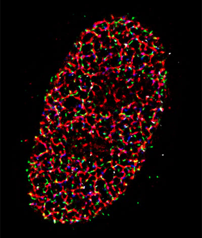
Image: STORM Super-Resolution image, taken with the Prime 95B Scientific CMOS camera. Cell Type: U2OS cells. Exposure time: 30 ms. Magnification: 150 times (configured pixel size ~ 73 nm). Reconstruction Algorithm Used: Maximum Likelihood Estimation (MLE) method for single PSF fitting. Image courtesy of Yandong Yin and Eli Rothenberg, New York University, School of Medicine.
At this point, Photometrics dominates the field with the only back-thinned sCMOS camera currently available, but that’s likely to change soon as other manufacturers enter this very competitive arena.
Biocompare’s Microscope Camera Search Tool
Find, compare and review microscope
cameras from different suppliers Search
But don’t count CCD out just yet. “CCD cameras are still a big part of microscopy imaging that are used on thousands of microscopes and many, many more are purchased each year,” notes Olenych. “They are the camera option that many people choose for their microscope imaging needs due to their relatively low cost and versatile performance.”
Additionally, for low-light imaging in spinning disk confocal microscopy or single-molecule studies, some users may still prefer the electron-multiplying CCD cameras, which use on-chip multiplication gain to achieve single-photon detection sensitivity without compromising QE or resolution. “This is a research-grade camera, used forever for these two applications. But honestly, even in these cases, I would argue that anyone thinking about buying a new camera should consider the new back-thinned sCMOS cameras,” Dr. Watkins suggests. “The only time you’d really want an EMCCD camera is when you absolutely need the amplification circuit, and that may contribute noise issues.”
In some circumstances, such as with an array-based detector on a confocal microscope, or when dealing with very fast low light, the user may still find that EMCCD is better. The only way to know for sure: get both cameras into the environment in which you’ll be using them, and test them. “That’s the primary message for buying a microscopy camera: test everything, buy once,” Dr. Watkins cautions. “I see a huge amount of buyer’s remorse with these cameras. Ask your vendor to let you test out the camera or cameras you’re considering in real-world lab conditions.”
After the great leap forward of backthinned scientific CMOS, what’s next for microscopy camera technology? “The race between manufacturers so far has been to increase the speed of the camera while at the same time increasing the QE,” says Claxton. “With QE for many of the sCMOS cameras now at 95%, you can’t go much further from there. As more back-illuminated sCMOS cameras are introduced, they will hit that limit. In the very near term, what we are likely to see is combining high QEs with low noise, while making the pixels smaller—a big field of view, but not sacrificing resolution to do that. The dream in the short term is to see things at a very high resolution, but over a very large area.”