A fluorophore is an organic molecule with the ability to absorb light at a particular wavelength and then emit it at a higher wavelength. To achieve this, photons of light from an excitation source are absorbed by the fluorophore’s electrons, raising their energy level and causing them to move to an excited state. To return to their ground state the electrons release light photons, yet since some of the excitation energy is dissipated, for example through transfer to neighboring molecules or via molecular collisions, the emitted photons have less energy and therefore a longer wavelength than those that were used for excitation; this allows the emitted fluorescence to be clearly distinguished from the excitation light source.
Fluorophores can be broadly categorized as organic dyes (e.g., fluorescein, rhodamine, AMCA), biological fluorophores (e.g., green fluorescent protein, phycoerythrin, allophycocyanin) and quantum dots. They are employed in a wide range of research applications including as antibody labels to indicate the presence of a target antigen in a sample via immunofluorescence, as reporter molecules when introducing a gene construct into a cell, as stains for the quantitative measurement of nucleic acids, and as indicators of cell health.
Fluorescent readouts are highly sensitive since the process of fluorescence is cyclical; each fluorophore possesses the ability to be repeatedly excited, resulting in the generation of a large number of detectable photons. Provided the fluorophore does not succumb to photobleaching—the phenomenon by which the ability to fluoresce is lost and which can occur, for example, through prolonged exposure to excitation light—samples can be read multiple times. Furthermore fluorescent readouts are ideally suited to quantitative analysis, and offer the opportunity for multiplexing, providing considerable savings in terms of time, money, and precious sample material.
How might you select a suitable fluorophore?
With such an extensive range of fluorophores commercially available the choice of a suitable molecule can be daunting, yet there are several key points to consider:
- The instrument that will be used to measure the fluorescent signal Fluorescent detection lends itself to a wide range of applications, yet invariably relies on a specialized reader for measurement of the signal output. Whether using a fluorometer, a flow cytometer, or a lateral flow strip reader, the dye that is chosen should be compatible with the excitation/emission channels of the instrument.
- The abundance of the target While some proteins are highly abundant in cells, others are present at only very low levels, and it can be beneficial to use a brighter fluorophore for detection of these targets, especially if the sample exhibits high background fluorescence. Another effective method for detecting low abundance targets is to employ a fluorophore-streptavidin conjugate to amplify the signal; by, for example, incubating the sample with such a conjugate following incubation with a biotinylated antibody or DNA probe, the signal output can be increased significantly.
- The excitation and emission spectra of the fluorophore The excitation and emission spectra of the fluorophore are of primary importance. Not only should they be carefully matched to the lasers and filter sets of the reader that will be used for detection, but they should also be compatible with any additional fluorophores employed within the experiment. Techniques such as flow cytometry and fluorescence in situ hybridization (FISH) often rely on multicolor labeling, and an ideal fluorophore combination for such applications would share the same excitation wavelength while demonstrating well-separated emission spectra to allow different readouts to be clearly distinguished from one another.
- The fluorophore’s Stokes shift The Stokes shift defines the difference between the maximum absorbance and emission wavelengths of the fluorophore, and the higher the value the greater the separation. This is of particular importance in multiplexing experiments, during which the emission of one fluorophore may overlap with the excitation of another, leading to unwanted background signal.
- The brightness of the fluorophore The fluorophore’s brightness is determined by its molar extinction coefficient and quantum yield; the former defines the amount of light that can be absorbed at a given wavelength and is measured in M-1 cm-1, while the latter is calculated through dividing the number of photons emitted by the fluorophore by the number of photons absorbed. Fluorophores with higher extinction coefficients and quantum yields are generally brighter.
- The cost of the fluorescent reagent Cost is a primary consideration in any experiment, particularly for those assays that will run for an extended period of time or that will be used to generate data from a large number of test samples. While novel dyes may be more expensive than those that have been in use for years, this is not always so, and significant cost savings can be made through bulk buying the fluorescent reagent or reserving batches of a specific lot for call off at a later date.
Designing a multicolor panel for flow cytometry
One application that relies heavily on multicolor fluorophore panels is flow cytometry, during which fluorescently labeled antibodies are used for the detection of specific proteins expressed by cells within a sample. One hurdle when building a panel of labeled antibodies is that researchers must often search multiple supplier companies for antibody/fluorophore combinations that are compatible with their flow cytometer. FluoroFinder says it has eliminated this problem.
Biocompare’s Flow Cytometry Search Tool
Find, compare and review cytometers
from different suppliers Search
“FluoroFinder specializes in cloud-based fluorescence experimental design tools to find the best combination of compatible fluorochromes for a flow cytometry or microscopy experiment,” explains Daren Young, director of business development. “We offer a comprehensive database of fluorochromes and instrument configurations that our interactive tools leverage to enable scientists to design and save complex multicolor experiments that are compatible on their instrument configuration of choice.”
Another company with expertise in multicolor panel design is BioLegend, whose diverse team of dedicated immunologists and advanced flow cytometry specialists have extensive knowledge of panel design and optimization. “Researchers can supply BioLegend with information including sample type and species, target antigen, preferred fluorophore, instrument model, and available lasers, and our specialists will then provide panel construction as a complimentary service,” explains Dzung Nguyen, director of marketing.
“We also provide online tools to help researchers design their own panels, such as our spectra analyzer, multicolor flow cytometry guide, and multicolor panel selector. BioLegend manufactures over 16,000 quality antibodies, and we offer the broadest selection of fluorochrome conjugates for multi-color flow cytometry. While many of our antibody conjugates are available as off-the-shelf products, should your desired antibody/fluorochrome combination not be available, we also provide an affordable, high-quality custom antibody conjugation service with rapid turnaround times.”
How critical are the emission filters to multicolor flow cytometry
Modern multicolor flow cytometers have the ability to simultaneously measure up to 20 distinct fluorophores, which places extremely high demands on the filters that are used to gather and differentiate the signals. Sarah Locknar, Ph.D., staff scientist and technical business development manager at Omega Optical, explains that “since the emission spectra of fluorescent dyes tend to be wide, there is considerable spectral overlap between adjacent dyes. This becomes more pronounced as the number of fluorescent labels is increased. The result of this overlap is that the signal collected at a particular channel will be a combination of the emission of the intended dye and significant emission contributions from adjacent dyes.”
Locknar expands further on this point, stating that “in multichannel systems it is essential that the spectral bandwidths of the emission filters are selected not only to optimize collection of the desired fluorescent signal, but also to minimize cross talk between channels and the need for color compensation. Compensation is a technique whereby interfering signals from other fluorescence channels are subtracted from the measured fluorescence signal, in essence removing crosstalk from the system.”
Omega Optical has been involved in fluorescent filter products since the technique of immunohistochemistry gained prominence during the 1980s, reportedly becoming the first company to offer multiband filters for simultaneous visualization of multiple chromophores. Since that time the company has continued to work closely with researchers in the microscopy and flow cytometry communities to establish specific band shape characteristics to match new dye offerings and that minimize the need for color compensation.
“By designing narrower pass bands and placing them optimally on emission peaks,” says Locknar, “we’ve reduced the relative contribution of an adjacent dye to a channel's signal, thereby producing a purer signal with less need for color compensation.”
The use of fluorescent dyes to quantify nucleic acid concentration
While many people automatically think of flow cytometry when they contemplate the use of fluorophores for research, the Promega QuantiFluor® dye systems for dsDNA, ssDNA, and RNA are a great example of harnessing fluorescent dyes to simply, quickly, and effectively quantify nucleic acid concentration.
“The QuantiFluor dyes have been specifically designed to preferentially bind a given species of nucleic acid,” explains Adam Blatter, Promega product specialist, clarifying that “when a dsDNA dye, for example, is excited by a given wavelength of light, only dye in the dsDNA-bound state will fluoresce.” As the dye binds its nucleic acid target, fluorescent quantum yield increases as a function of shift in fluorophore molecular geometry. “This aspect of the QuantiFluor dyes, in conjunction with their binding to specific nucleic acid species, contributes to a low background signal, high accuracy, and specificity,” says Blatter, “making them ideal for low-level nucleic acid quantitation from FFPE tissue, ccfDNA, and other similar sample types.”
As with any fluorophore, the QuantiFluor dyes must be used in conjunction with fluorometers equipped with the correct excitation/emission channels, but provided this is the case QuantiFluor dye systems can easily be scaled in throughput from a single sample at a time to 96- and 384-well plate formats.
“The Promega Quantus Fluorometer is designed to provide convenient and highly sensitive fluorescent detection for a single tube per read using the two channels required for this specific application,” adds Blatter. “For labs requiring a higher-throughput plate-based fluorescent quantitation method, or more filters for additional applications, then multimode readers such as the Promega GloMax Systems may be used.”
What if the antibody/fluorophore combination isn’t available?
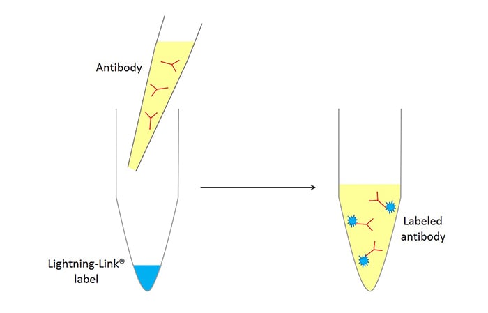
With such a huge array of antibodies available, it is often the case that the desired antibody/fluorophore combination isn’t available as an off-the-shelf product, since for manufacturers to provide every possible option they would have to stock an almost infinite number of products. To overcome this Innova Biosciences offers Lightning-Link®, antibody labeling kits that allow the end user to directly conjugate their antibody to a fluorophore with only 30 seconds hands-on time and 100% recovery of materials.
According to Anna Sereni, head of research and development, Lightning-Link works by targeting free amine groups within the antibody to attach the fluorophore label via a covalent bond. “We offer almost forty different fluorescent labels within the Lightning-Link range, covering the spectrum from ultraviolet through to infra-red”, she says, “and this includes fluorescent dyes, fluorescent proteins, and also tandem labels.”
Tandem dyes can be used to increase the quantity of readouts from an experiment should the number of excitation lasers be a limiting factor; for example, DyLight® 488, R-phycoerythrin (PE) and PerCP/Cy5.5 can all be excited using a 488 nm laser, yet generate green, orange, and red emissions, respectively. Sereni adds that “our Lightning-Link technology is fully scalable, allowing you to label as little as 10 µg antibody up to 1 mg or more, while our stringent QC process provides consistent high quality and excellent batch-to-batch reproducibility.”
 The Lightning-Link products have been cited in over 500 publications to date, and Innova Biosciences also offers accessory kits to ensure that the antibody is in a favorable starting position for labeling (correct concentration, removal of interfering buffer components), as well as kits to quickly and easily confirm that the conjugation was successful.
Image: Schematic representation of the Lightning-Link® conjugation process. Image courtesy of Innova Biosciences
Summary
The choice of fluorophore is dependent on the type of experiment that will be performed, as well as the instrument that will be used to measure the fluorescent signal, yet despite the fact that it is theoretically possible to exchange one fluorophore for another, this is not always the case in practice. Although two fluorophores may share the same maximum excitation and emission wavelengths, factors such as the brightness of the dye and the width of the emission spectra should be given due consideration, especially when implementing a multicolor panel.
While the range of commercially available fluorophore products is extensive, and continues to grow, the services offered by companies such as FluoroFinder and BioLegend greatly increase the ease of navigating these reagents. Vendors such as Innova Biosciences provide a neat solution in situations where the desired antibody/fluorophore combination is unavailable, while others offer expertise in the development of new dyes, the provision of specialized readers, and the production of fluorescent filters to improve data quality through the generation of purer signals. Fluorescent detection is extremely sensitive, applicable to a wide variety of techniques, and is ideally suited to multiplexing experiments; with continuing advances in fluorescent reagents and reader technologies, fluorophores have an incredibly bright future—no pun intended!