ReCyte Therapeutics, A Subsidiary of BioTime
Associate Scientist
Angiogenesis, or neovascularization, is the process of generating new blood vessels from existing vasculature. In vivo, endothelial cells (ECs) must migrate through the basement membrane, proliferate and form tubular structures. In vitro, ECs seeded onto the extracellular matrix substrate Matrigel mimic the late stages of angiogenesis and form tube-like structures. The in vitro Matrigel tube-formation assay has been used extensively to screen for the angiogenic activity of hundreds of chemical compounds and biologics. The traditional in vitro Matrigel tube-formation assay is robust and scalable; however, it is limited by poor tube stability after one day of cell culture and therefore limits the ability of researchers to study multiple angiogenic events.
Co-culture solutions
To address the limitation of tube stability, in vitro co-cultures of ECs with mesenchymal stromal cells (MSCs) or perivascular cells (PCs) have been developed to prolong the lifetime of tube networks generated by ECs, including human umbilical vein endothelial cells (HUVECs). Co-cultures containing MSCs utilize these cells as a feeder layer. To generate a feeder layer, hundreds of thousands of MSCs are seeded on tissue-culture plastic and form a dense monolayer. HUVECs are then seeded onto the feeder layer and HUVEC tube networks form within one day. Notably, these tube structures mimic in vivo capillary formation and are stable over multiple days. However, the feeder layer cells show little to no integration with EC tube-like structures and further require the addition of exogenous growth factors to support long-term stability. Alternatively, co-cultures containing PCs use these cells to direct and stabilize EC tube-like structures on Matrigel. The resulting co-cultures show rapid tube formation within four to eight hours, comparable to HUVEC monocultures on Matrigel. The addition of PCs results in enhanced tube stability for at least four days without the need for exogenous growth factors. In normal physiology, PCs are essential for the maturation and stabilization of vasculature, and their dysfunction is associated in a variety of physiological disorders (e.g., tumor progression, stroke and macular degeneration). In an in vitro co-culture, PCs stabilize HUVEC tube networks via both direct cell-cell contacts and paracrine signaling pathways. Therefore, PC-based co-cultures have the significant advantage of providing a physiologically relevant tube-formation and stability assay. Moreover, the ability to use basal media during the assay enables easy quantification of the effects of test reagents on the co-culture.
Selection of PCs for co-culture assay
There are multiple sources for PCs that can stabilize EC tube networks in co-culture. For in vitro co-cultures, the following PC types are suitable for co-culture with HUVECs: CD146+ bone marrow-derived MSCs (BM-MSCs), CD146+ adipose-derived stromal cells (ADSCs), placental-derived pericytes (Pl-PCs), brain vasculature-derived pericytes (BV-PCs), and a pericyte-like progenitor cell line (PC-M). The use of primary cell populations is advantageous for in vitro assays requiring elucidation of the interactions between endogenous cell types and may best mimic the in vivo environment. However, the use of primary cells is prone to batch to batch variability. In contrast, PC-M cells are derived from the human embryonic stem cell (hESC) line ESI-017 and show stable marker expression of CD146 over 90 population doublings. In vitro, PC-M cells stabilize HUVEC tube networks for longer time periods than observed in co-cultures containing BM-MSCs, Pl-PCs or BV-PCs. Therefore, the use of PC-M cells instead of primary cells supports greater stability and reproducibility in an in vitro angiogenesis co-culture assay.
Optimizing culture conditions
PC co-culture assays can be optimized by further altering the total cell-seeding density, the ratio of PCs to HUVECs and the density or type of substrate. A range of cell densities and ratios have been reported for co-cultures containing PCs and ECs. Here are some tips on selecting the optimal experimental conditions to use.
To observe the stabilizing effects of PCs, the EC seeding density must correspond to EC monocultures that form tube networks and then degrade in the absence of PCs. This occurs at HUVEC cell densities on the order of 1 to 1.4 × 10⁵ HUVECs per cm2. To observe the stabilizing effects of PCs, a ratio of 1:3 to 1:50 PCs to HUVECs may be used. Depending on the type of vasculature and stage of development, the ratio of PCs to HUVECs also varies greatly in vivo and thus is in an important variable in any co-culture assay. For in vitro angiogenesis assays on Matrigel, we have found that ratios of 1:5 to 1:20 (PCs to ECs) enhance the stabilizing effects of BM-MSCs, Pl-PCs and PC-M cells.
Finally, the selection and density of substrate can alter the ability of cells to migrate and co-localize. Two commonly used substrates, Collagen Type I and Matrigel, promote tube formation at concentrations of 2.5 to 3.75 mg/ml and 9.0 to 10.0 mg/ml, respectively. The volume of substrate required depends on the well size. For example, substrate volumes of 50 to 80 µl per well are used in a 96-well plate assay.
Testing pro- and anti-angiogenic compounds
PC co-culture assays can be used to test the effects of multiple pro- or anti-angiogenic compounds at multiple time points. Here are some considerations when setting up an analysis.
Test reagents should be added to the assay culture following initial tube formation, which occurs as early as four hours after initial cell seeding. The tube formation at early time points and in untreated cultures are both used as reference points. When testing pro-angiogenic compounds, researchers should observe increased tube length and branching, as well as enhanced stability over time. When testing anti-angiogenic compounds, researchers should observe decreased tube length and branching, as well as tube instability over time. Further optimization of the test reagent dose and repeat doses (e.g., timing of delivery) is easy to accomplish in one assay, because tube-like structures in untreated reference co-cultures are present for at least four days. The successful completion of any PC-based co-culture assay requires an assay system that includes: (1) PCs that make direct cell-cell contacts with ECs, (2) long-term tube stability and (3) the absence of exogenous growth factors for easy analysis.
The VascuNetTM Pericyte Co-culture Assay from ESI BIO can be used to establish extensive HUVEC tube networks that are stabilized by PC-M cells for at least four days of cell culture. PC-M cells utilized for the VascuNet Pericyte Co-culture Assay co-localize along the entire length of tube-like structures (Figure 1). The VascuNet Pericyte Co-culture Assay thus presents a more physiologically relevant model of tube formation and stability for scalable screening of pro- and anti-angiogenic factors, as well as their long-term effects on tube formation.
Following these tips and suggestions on PC-selection options and seeding-cell densities in assay conditions will enable you to overcome the challenges of tube stability when performing in vitro angiogenesis experiments.
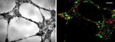
Figure 1: Bright field (left) and fluorescent (right) images of representative tube formation following co-culture of PC-M cells (red) with HUVECs (green) at Day 1. Cells were seeded at 42,000 cells per well on 96 well plate coated with Growth Factor Reduced Matrigel®. 4X magnification; scale bar=150 mm.
Related Products from: ESI BIO-A Dvision of BioTime, Inc