The complexity of biology often demands the combination of techniques to really unravel processes. One of the most powerful technological blends is immunohistochemistry (IHC), which combines anatomical, biochemical, and immunological techniques. Scientists use IHC in basic biological research and in diagnosing diseases and developing drugs. Getting more out of IHC requires ongoing improvements in product chemistries.
In 1942, American immunologist Albert Coons and his colleagues described IHC in The Journal of Immunology. As the scientists wrote: “The object of this investigation has been to determine the feasibility of using chemically labeled antibodies as reagents for the detection and orientation of antigenic material in mammalian tissue.” And, feasible it was. In fact, scientists around the world use IHC as a routine method.
According to an overview of IHC from Thermo Fisher Scientific: “IHC makes it possible to visualize and document the high-resolution distribution and localization of specific cellular components within cells and within their proper histological context.”
Many scientists and clinicians gain insight from IHC. For instance, Michael Leapman of the Yale School of Medicine, says, “As a urologist who treats patients with genitourinary cancers, I rely on IHC analysis in pathology reports to help make diagnosis and guide treatment and clinical management.” He adds, “For example, numerous IHC markers are used for the diagnosis of kidney cancers, including cytokeratins, vimentin, PAX2, PAX8, CD117, TFE3, III, p63, CD57, carbonic anhydrase IX, etcetera.” The use of IHC in medicine involves many specialties. For example, Leapman says, “As a surgeon, I do not use the markers directly in clinical care but rather rely on their interpretation and performance by pathologists.”
With so many uses of IHC, many people—from basic researchers through clinicians—must know how to use the method or interpret the results.
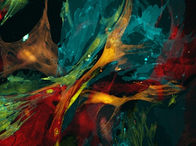
Image: Immunohistochemistry can be used in a range of basic research and clinical applications, such as treating kidney cancer. In these kidney cells from a so-called ‘confetti mouse’, antibodies light up features based on molecular differences, such as characteristics that distinguish tumors. Image courtesy of Heinz Baumann, Sean T. Glenn, Mary Kay Ellsworth, and Kenneth W. Gross, Roswell Park Cancer Institute, Buffalo, NY.
Overcoming obstacles
IHC involves various reagents and tools. As Sino Biological describes it: “Immunohistochemistry is a technique that uses antibodies (matching molecules) that can seek out, identify, and attach themselves to these markers on cells. The antibodies themselves can be seen under the microscope, which helps the technician make a precise identification.” The processes involved require skills in a collection of techniques and familiarity with many products. For example, Sino Biological notes that it “has launched nearly 1000 immunohistochemical antibodies.”
Despite the broad use of IHC in labs around the world, all scientists need a place to start with this technique, and ways to work around problems. Boster Biological Technology provides a detailed introduction to IHC, which goes from sample preparation to detection.
Plus, Enzo Life Sciences developed 10 Tips for Successful Immunohistochemistry, which explores tissue preparation and fixation, blocking non-specific binding sites, selecting a detection system, and more. As an example, this guide notes: “When multiplexing your IHC staining with different markers and consequently different chromogens, you should keep in mind that your counterstain will need to stand out and provide true background contrast.”
Sometimes, though, IHC doesn’t work as well as expected. For such stumbling points, Thermo Fisher Scientific runs an online IHC Troubleshooting Guide, which it states: “discusses the major problem areas in IHC staining.”
If sitting back and watching feels like a better way to get started with IHC, take a look at the Basics of Immunohistochemistry. In this webinar, Jeff Gordon, OEM sales manager for tissue diagnostics at MilliporeSigma, describes the basics of immunohistochemical techniques, the science behind IHC, different detection methods, and the monoclonal and polyclonal options.
Such online tools and discussions with colleagues help scientists get started and solve many problems in applying IHC.
Adding enhancers
The steps and results in IHC depend fundamentally on the reagents used. Some of those are designed to provide more information. For example, Edward Rosen, vice president and managing director at Cosmo Bio USA, describes immunoreaction enhancers (IEs) for IHC as “a class of reagents that offer improved performance in any antibody-based assay system, including with frozen and paraffin-embedded tissues.” He adds, “IEs are proprietary solutions of polymers and other substances offered by several makers that are most often provided as ready-to-use solutions to replace existing antibody diluents.”
As one example, Rosen mentions the Can Get Signal Immunostain Immunoreaction Enhancer, which is made by Toyobo and made available worldwide from Cosmo Bio. “Toyobo offers IEs formulated specifically for primary or secondary antibodies,” Rosen says. “IHC experiments employing Can Get Signal solutions may significantly reduce background staining and improve antibody sensitivity and specificity to boost signal-to-noise ratios.” He adds that this product “is compatible with ABC and polymer-based detection kits.”
The Can Get Signal product offers a range of other useful features. For one thing, it can increases sensitivity and specificity in IHC applications. As well, this IE can be used, Rosen says, with “various detection systems—for example, chromogenic, chemiluminescence, or fluorescence.”
Lab applications of IHC
Scientists use IHC to study a range of biological processes. Take autophagy, for example. This process performs many tasks, such as destroying damaged organelles and proteins in cells. Some scientists describe it as molecular housekeeping. It could also play a part in preventing cancer, and possibly various degenerative diseases.
At Istanbul University in Turkey, Melek Öztürk and her colleagues use IHC to study autophagy-related genes that play a role in cell death. As these scientists noted: autophagy-related genes regulate autophagy and “control the crosstalk with autophagy-associated cell death and apoptosis” in some conditions. Although scientists can use quantitative real-time PCR to study the expression of these genes, Öztürk and her colleagues pointed out that the “detection of the markers for autophagy-related process[es] by immunohistochemistry in paraffin sections of various patient tissues has become a reliable method for monitoring autophagy.”
That’s just one of many ways that IHC can be used in the lab. In fact, many users—scientists in an academic lab or industrial engineers— find ways to use IHC. In all cases, Coon would surely be pleased to see the breadth and benefits of his work.