Cells and their culture are integral to biomedical research. The ability to reliably and efficiently assess their wellness is a critical aspect of high-quality and reproducible experimentation. Cell health measurements are used for studying the effects of drugs, cell culture monitoring and optimization, assessing stem cell differentiation, and more. With so much riding on cell health assessments, getting it right is pretty important. So we asked six experts from companies that know a lot about cell health to share their knowledge with us to help ensure your cells and cultures are healthy and can help you achieve reliable and reproducible experimental results.
Our expert panelist include: Mike Blundell, Ph.D, Product Manager at Bio-Rad; Klaus Bischoff, Head of Research Solutions at MilliporeSigma; Pam Guthmiller, Director, Product Management, Cellular & Protein Analysis, Promega; Tracy Murphy, Director of R&D and Quality Control, ImmmunoChemistry Technologies (ICT); Robert Newman, Senior Director at ATCC Cell Systems; and Kalpana Patel, R&D Scientist at Essen BioScience (Sartorius).
Their insights, tool and methodology suggestions, and best practices are included in the following discussion.
1. Can you weigh in with your thoughts on the importance of assessing cell health?
Blundell: For many experiments, researchers need to use healthy cells so they can accurately assess cell responses, treatments, and survival. If a cell population is unhealthy from the start of an experiment, or if the cells’ health status is unknown, it is difficult to assess the significance of any changes observed in these cells during the experiment.
Cell health must be measured thoroughly using multiple criteria during an experiment to ensure the results will be reproducible. Cell viability is often one of the critical measurements, but it is not the only indicator of cell health. Cell health can also be determined by measuring proliferation responses and the progression through apoptosis and autophagy.
Isolating healthy cells is important, for example, in flow cytometry, where a clean signal is key. When staining cells with antibodies for a flow cytometry experiment, the antibodies can bind to dead cells nonspecifically, leading to increased background noise. This background noise can not only produce false positives, but can also reduce the resolution of the signal in multicolor panels. Color can spill over and fluorescence can spread, obscuring the presence of dim fluorescent samples.
It is also important to measure cell health when measuring apoptosis using annexin and propium iodide (PI) staining. If the health status of a cell population is unknown before an experiment starts, it is difficult to assess the effect of a particular treatment on these cells. If detects an increase in fluorescence is detected using these stains, it will be unclear whether it is because the unhealthy cells in the population are reacting differently, wherein the kinetics of apoptosis have changed.
Bischoff: Carrying assays out on cells whose viability or proliferation is not detected beforehand can lead to erroneous results and can be very costly to the researcher. For example, if cells are either dying or dead before performing compound treatments, cells will be more likely to show induction effects that are not specific to the assay being evaluated. Additionally, if cells are dead or dying when performing studies with fixation and permeabilization protocols, large cell loss can be observed.
Guthmiller: Good assay results are highly dependent on the health of the cells you start with. There are many factors to be aware of when handling cells and preparing them for an experiment. Many of these activities can reduce drift in the phenotype of cells, which can result in lack of reproducibility. Defining standard operating procedures in your lab for banking and maintaining cells in culture and handling cells for assay implementation is highly recommended. These should include developing cell banks, contamination testing, cell line authentication, documenting passage number when working with stably transfected cell lines (which can change expression levels from extensive passaging), and determining appropriate plating cell numbers for the treatment time desired to minimize adverse effects of overcrowding such as contact inhibition. If cells are being used to express proteins, understanding the appropriate culture conditions is imperative for optimal expression. A good source of basic information is R. Ian Freshney’s Culture of Animal Cells: A Manual of Basic Technique and Specialized Applications, sixth edition (Wiley).
Murphy: Given the time and expense of cell culture–based research, it’s critical that cell cultures used for experimental studies are properly handled and maintained. When unhealthy cells are used, the potential for experimental error or artifacts is increased. For example, cells in a suboptimal state of health may have lost or reduced ability to respond normally to a particular experimental treatment. Poor cell culture conditions may also lead to proliferation of a particular genetic clonal subset in what is supposed to be a genotypically identical population. Last, suboptimal cell culturing practices contribute to high levels of spontaneous apoptosis or dead/dying cells within the population. Given these factors, experimental conclusions drawn from studies using unhealthy cells are clearly problematic at best.
Newman: For reliable and reproducible cell culture–based studies results, it is important to ensure that you have control over your cell culture process to determine that your material is consistently of the same quality. Cell health can be measured during expansion of cell banks, prior to initiating cell-based studies, and during in-process time points or at the end of studies. It is important to measure cell health when cell viability is critical, or when you are testing specific endpoints, such as apoptosis, metabolism, cells stress, and toxicity.
Patel: Although obvious, it is worth stating that the cells used within cell-based models are critical to the success of the assay. Making noninvasive assessment of the cells throughout the continuum of cell culture, manipulation, and assay can provide a measure of growth quantification and a qualitative record of morphology. These, in turn, can be related to the study output to better understand the causes of spurious results, thus improving reproducibility.
 Records of proliferation and associated morphological images provide a reference to enable appreciation of changes of time (weeks, months, or years) where cell types may have differentiated following excessive passage. With a greater emphasis on translational models, the cell types employed are becoming more challenging—whether induced pluripotent stem cells (iPSC)-derived or patient-specific—therefore, the need to monitor the health and growth profiles of these highly valuable cells is elevated. Overall, the boundary between cell husbandry and assay is becoming more blurred.
2. What are the critical parameters to measure to determine cell health?
Blundell: Critical parameters include: (1) Cell viability. This can be measured using either cell-impermeable DNA-binding dye such as PI or 7-aminoactinomycin D (7-AAD), or protein-binding dyes that one can fix for convenience. (2) Proliferation responses. Even if a cell appears healthy and is viable, it may not be able to proliferate, which may affect the results of an experiment. (3) Apoptosis and autophagy levels. These metrics allow one to detect initial signs of cell death or cellular stress in otherwise healthy-looking cells.
Bischoff: Traditionally, cell viability and concentration have been the hallmarks of cell health, and they still are, but this view has changed in recent years. It is being recognized that there are other critical parameters. Quick determination of both cell concentration and viability and their assessment over time, as well as culture conditions, are baseline data that researchers must obtain regularly. But periodic assessment of apoptosis and mitochondrial perturbations can provide earlier and more sensitive indicators of cell health changes.
Guthmiller: Cell health can be monitored using a variety of biomarkers. The choice of methods is dependent on what question you are trying to answer. If you just need to know how many cells per milliliter of culture you have, a simple method to count cells with a stain, such as trypan blue, is sufficient. However, if you want to know more specifically how cell culture conditions or treatments are affecting your cells, there are numerous options to assess viable cells, dead cells, or dying cells.
The two most common methods to assess viable cells are by measuring the metabolic capacity of cells either by measuring their capacity to reduce a substrate or by measuring changes in adenosine triphosphate (ATP) content. Methods to detect dead cells determine whether the cell membrane is intact and can be measured with dyes that only get into cells with compromised membranes or by detecting proteins that leak out into the cell culture medium when the membrane is compromised, such as with the lactate dehydrogenase (LDH) release assay. To assess whether cells are under stress, reactive oxygen species (ROS) can be measured. Several markers that are used to assess whether cell signaling events have triggered cells to die through the mechanism of apoptosis include detection of caspase-3/7 activation or the flipping of phosphatidylserine to the outer cell membrane.
Murphy: Some of the parameters we monitor in our lab are growth rate (doubling time), morphology (size/shape/appearance under the microscope), concentration (3 x 10⁵ cells/mL –7 x 10⁵ cells/mL is generally ideal for suspension cell lines). We also can tell quite a bit by looking at the forward versus side scatter (FSC vs. SSC) on flow cytometry density plots. Healthy cell populations should have relatively uniform FSC vs. SSC characteristics, without evidence of excess cellular debris/smaller particulates within the population.
Live/dead stains, such as 7-AAD, PI, and Green Live/Dead Stain, provide a quick and easy quantitative method of measuring cell viability using flow cytometry, and can deliver results in under 10 minutes. If time allows, detection of caspase activity using FLICA® (alone or in combination with a live/dead stain) can provide a more detailed picture of the overall health of the cell population. This is because caspase activation occurs very early in the apoptotic pathway, before the cell membrane becomes compromised (when cells would begin to stain positive for a live/dead stain).
In cases where the symptomology is more subtle, but nonetheless capable of compromising cell performance leading to experimental artifacts, it might be helpful to assess the general mitochondrial polarization status of your cell culture stocks. Cell culture media–associated nutritional factors as well as excess numbers of cell passages can commonly play a role in the degradation of cell health status. Events such as these do not necessarily lead to an immediate conversion to an apoptotic or necrotic state. They might very well manifest themselves in the form of a generalized reduction in mitochondrial polarization status. Employment of a variety of fluorescent potentiometric dye products could identify these underlying problems in advance of committing a lot of time and energy setting up and carrying out your particular experiment.
Newman: Critical parameters for cell health include cell viability and phenotype indicators, such as morphology, cell marker expression, and cell biofunctionality. Depending on your objective, other critical parameters could be testing for metabolites, apoptotic markers, stress signals, toxicity markers, or indicators of cell lysis.
Patel: Cell health is often determined by assessing the proportion of dead cells using membrane integrity assays. These include PI and trypan blue exclusion assays and provide a robust measure of percent viability. However, a more reliable approach is to assess the health of the living cells. Growth rates can provide a more robust measure of cell health, notably enabling subtle changes that reduce proliferation without inducing death. Cellular metabolic activity (LDH, ATP, MTT, alamarBlue®) can be used as a surrogate for cell number; however, this only provides information at a single time point and may be influenced by changes in metabolic rates. Measuring the full time course of cell proliferation using live cell analysis techniques can provide an insight into cell health. Unhealthy cells are likely to exhibit altered proliferation rates compared to their healthy counterparts. Additionally, live cell analysis offers chronological records of cell morphology, which can be used to identify changes immediately and historically.
3. How often should cell health be assessed and are there specific events or actions that should trigger testing?
Blundell: Cell lines should be assessed regularly for cell health. In fact, testing a cell population’s viability after each passage serves two purposes: (1) It indicates cell health, and (2) it allows one to accurately seed the cells at the right concentration, in preparation for the next experiment.
Cell health should also be tested prior to the start of an experiment. As mentioned previously, cell health data should be included in your experiment to enable one to assess whether any observed changes in the cell population during the experiment were due to changes in the dead cells.
Bischoff: Assessment of cell health is dependent on the cells being evaluated and specific lab protocols. Generally speaking, determination of the cellular concentration and viability should be done directly prior to splitting or passaging the cells. Monitoring cell health when cultures are aging is also important, since characteristics of cell lines can change as they age. If cells are being used for assay purposes, it is good to evaluate the cellular concentration and viability both the day before assaying as well as the day of.
Guthmiller: The health of your cells should always be assessed whenever you do any manipulations, such as splitting, passaging, transfection, changing culture conditions, or any type of treatment. The first and most obvious test is simple morphological observation using an inverted microscope.
Murphy: Many of the above parameters (growth, morphology, maintenance at adequate cell concentrations) are easily monitored and should be done on a daily/ongoing basis. More involved testing, such as use of live/dead stains or fluorochrome inhibitor of caspases, or FLICA, testing, should be considered before an important experiment, or if other parameters (growth rate, etc.) seem off or indicate something of concern. Some of this will depend on the cell line or type, as some are more sensitive than others.
Newman: The frequency of cell health testing depends on the goals of testing. Cell health can be used as an endpoint measure or it could be used as an intermediate testing point during cell culture. Your objective will determine how often cell health should be measured. Sometimes cell health testing should be added for troubleshooting purposes. For example, if you start noticing variability in your experiments and there are indications of changes in cell viability or phenotype, you could try investigating cell health at in-process time points to determine if there are any issues with your cell culture process.
Patel: Traditionally, percentage cell viability achieved using trypan blue when cell counting at the point of cell passaging or seeding has been used as a surrogate of cell health. Although useful, this approach only provides a crude measure of the proportion of live/dead cells at a single time point. More often, the time when the researcher most wants to understand the health of cells is when the assay has underperformed. Invariably, it is too late to go back and fully quantify the cell’s health; however, if this becomes part of the routine culture process, then that would help.
 In addition, factors affecting assay performance may not manifest as cell death. Ability to appreciate the morphology of the cells both before and during the assay may aid the interpretation of the results. There is no fixed rule as to how often to determine cell health; however, whenever significant events occur, such as cell seeding, then assessment is recommended. If the cell type is unfamiliar and/or liable to differentiate, such as primary cultures, then more regular assessments should be made. Live cell analysis offers the opportunity to make noninvasive assessments, and thus can become part of the standard culture workflow.
4. Can you share any tips, products, or best practices to accurately and efficiently assess cell health?
Blundell: The easiest way to assess the general health of a cell population is to use a viability dye, which is an essential control for most experiments. For example, if a cell isn’t healthy and is unable to respond to a stimulus, it may be difficult to assess proliferation or cytokine release. For flow cytometry, researchers should include these dyes in every experiment, especially when using multicolor panels.
It is easiest to use nonfixable DNA-binding dyes such as PI, 7-AAD, or 4',6-diamidino-2-phenylindole (DAPI). To use a viability dye, simply add it to your cell suspension and analyze: Live cells with intact membranes will not fluoresce. But these dyes do have a downside—they cannot be fixed, and the space into which they can be inserted into a multicolor panel is limited.
But fixable viability dyes, such as in the VivaFix™ Cell Viability Assay, are available. In fact, they come in a wide range of excitation and emission spectra, and they are suitable for most instrumentation, giving you more flexibility in your experiments. These dyes can also be used in immunofluorescence microscopy.
When using reagents, ensure you follow the manufacturer’s instructions regarding the use of correct buffers. Additionally, you may need to optimize your protocols and titrate the reagent. If the reagent is present at too high a concentration, it may actually harm the cells and yield false results. It is also important to understand the configuration of your cytometer or your microscope and the choice of the most appropriate reagent.
Don’t forget to include controls. If you are performing multicolor experiments, use compensation controls. You may need to use both positive and negative controls to see the full range of cellular responses. As a negative control, include the vehicle used to resuspend the reagents.
Also, ensure you are using the right reagent for your experiment. There are many ways to measure apoptosis, for example. If you are unsure how a pathway works or if you do not want to measure specific components of the pathway, use a more general reagent. Caspase FLICA™ Apoptosis Detection Kits, for instance, allow one to detect various active caspases in virtually all mammalian cells, and they are available in four different formats, suitable for flow cytometry and microscopy. But the Caspase-9 FLICA Kit is only suitable for a specific stage in apoptosis: It detects progression through the intrinsic pathway only after mitochondrial membrane depolarization. A Poly Caspase FLICA Kit, on the other hand, will allow you to detect caspases involved at all stages of apoptosis.
Finally, before you start your experiments, ensure your samples are well prepared and carefully treated; keep them at the right temperature and concentration. This protocol will optimize the health of your cells, reducing the impact of poor cell health on your experiment. This can be especially important in applications such as Western blotting, where you cannot exclude dead cells from your analysis. You should still include reagents to assess cell health, however, where possible.
Bischoff: MilliporeSigma offers easy-to-use assays for both count and viability and annexin evaluation in a mix-and-read format for a variety of cells. Our Muse® Cell Analyzer and our Guava® easyCyte instrument offer assays with mix-and-read formats with as little as 10 μL of cellular samples and instructions that walk the user through the procedure for evaluating cellular health. The Muse Cell Analyzer is a closed platform with 25 optimized assays spanning various areas of cell health, while the Guava system is an open system with up to three lasers that is available in either a single- tube or plate-based format.
Guthmiller: The best method to choose for assessing cell health is dependent on many factors, including what you are trying to measure, what instrumentation you have available and your expertise to use the instrumentation, the type of samples you have, and the throughput desired. Many commercial assays are available to assess cell health in microwell plate-based formats. For simple yes/no experiments to assess whether a treatment affects cell health, often tetrazolium (MTT, MTS) or resazurin dyes that use cellular metabolism to convert them to colored or fluorescent products that are detected with a plate reader are implemented. However, these assay methods: (1) can contribute to toxicity from the detection reagent itself, (2) are not sufficiently sensitive for higher throughput of samples, and (3) are prone to interferences that make them less sensitive or result in false results.
For critical applications where precious samples are used and quantitation is critical, or when efficiency is needed, such as in higher-throughput experiments, assays that detect ATP are easiest to use because they follow a simple add–mix–measure protocol that provides data in as little as 10 minutes. Also, they are significantly more sensitive, thus requiring fewer cells and allowing them to be miniaturized for high-throughput applications. To optimize culture conditions or the effect of treatments, it is critical to understand the progression of changes in cell health over time.
With new real-time assays available for monitoring viability, cytotoxicity, or apoptosis over time from the same sample wells, such as with the new RealTime-Glo™ Viability Assay or the RealTime-Glo Annexin V Apoptosis and Necrosis Assay, you can now easily identify optimal parameters for growing cells or determining optimal treatment times and concentrations. Monitoring changes in the concentration of available nutrients in the culture medium and the metabolites produced by cells can offer insight into metabolic changes over time, such as switching from mitochondrial oxidative phosphorylation to glycolysis as the major source of energy production for cells.
Murphy: ICT’s Necrosis vs. Apoptosis Assay provides a convenient method for simultaneous detection of apoptosis and necrosis within a cell population using flow cytometry or fluorescence microscopy. This kit combines a red fluorescing live/dead stain, 7-AAD, with a green fluorescing reagent, FAM-FLICA, for caspase detection in a simple, easy-to-use test. With this assay, apoptotic cells stain positive (green) for FAM-FLICA, necrotic cells stain positive (red) for 7-AAD, and late-stage apoptotic cells stain positive for both FAM-FLICA and 7-AAD.
In addition to monitoring cellular health status in populations in ongoing cell culture–based research, the Necrosis vs Apoptosis Assay has utility for assessing cellular cytotoxicity properties of various chemical and biological agents; therefore, it provides an early screening option for safety studies associated with potential pharmaceutical agents, food additives, etc.
Should you be concerned about more subtle health status problems that don’t quickly evolve into apoptotic or necrotic cell death states, then ICT’s JC-1 or tetramethylrhodamine methyl ester and tetramethylrhodamine ethyl ester (TMRM/TMRE) fluorescent potentiometric mitochondria polarization status kits could be your solution.
Newman: To accurately and efficiently assess cells, you should have a very good understanding about what you are trying to measure so that you select the right assay (e.g., are you trying to test viability, metabolism, apoptosis, toxicity, lysis, etc.?). Next, you should ensure that you are using the correct cell type and model system (e.g., 2D or 3D culture). You should also evaluate the format of the assay (e.g., is it low throughput or high throughput?), the time course of the assay, dosing (if you are testing a treatment), and whether you desire an endpoint assay or a real-time nondestructive assay that does not kill the cells. You should also take into account whether you are trying to multiplex with other assays. Other standard assay considerations include evaluating ease of use, sensitivity, specificity, accuracy, detection limit and range, cost, and reproducibility.
Patel: Getting to know your cells is key. Regular visual inspection of the shape and appearance of your cells in culture and just before use will allow early detection of unhealthy cultures and ensure healthy cells are used for assay. Employing cell culture best practices can avoid cells becoming unhealthy. Aseptic techniques minimize culture contamination and optimal subculturing (splitting, passaging) at defined cell confluences can mitigate the build-up of metabolic waste and nutrient depletion in culture medium.
Avoiding assumptions around growth conditions and ideally using optimized media, supplements, and serum can further enhance cell health. Utilizing microtiter-based matrix experiments of media constituents allows for rapid and simple characterization of culture media, which is paramount when using valuable (e.g., patient specific or iPSC- derived) cells.
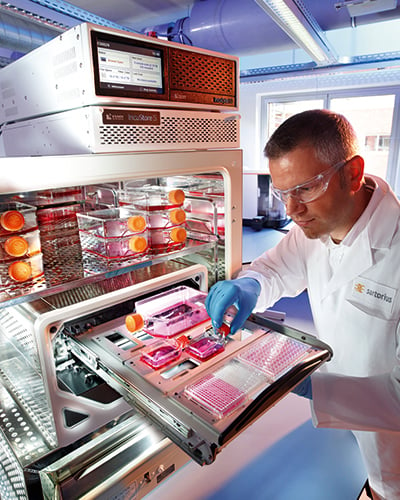
Image 1: Courtesy of Essen Bioscience.
Image 2: Courtesy of Dreamstime