Interest in the use of three-dimensional (3D) cell culture systems has grown significantly in recent years. Affording greater physiological relevance than traditional two-dimensional cultures, along with more predictive data for in vivo experimentation, a range of enabling technologies has been developed to improve understanding of a multitude of cellular complexities. These include highly specialized reagents and consumables for easier and more accurate visualization of cells in three dimensions.
…It is now possible to detect every single cell within a 3D cell culture model using confocal imaging with a high-content instrument.
According to Michael Johnson, CEO at Visikol, one of the major challenges that researchers encounter when attempting to study cells in 3D is that beyond 1 to 3 cell layers, 3D cell culture models cannot be effectively imaged. “The majority of confocal and widefield imaging assays are biased to characterizing only the periphery of 3D models, where cell proliferation and exposure to a compound or antibody is greatest,” he notes. “This can result in misleading insights, but by rendering a 3D model transparent using the Visikol® HISTO-M™ tissue clearing technology it is now possible to detect every single cell within a 3D cell culture model using confocal imaging with a high-content instrument.”
Visikol HISTO-M renders 3D cell cultures transparent through the process of refractive index matching, during which water is replaced with a high-refractive index solution. “This non-destructive reagent is fully compatible with immunodetection,” adds Johnson, “and using it we’ve observed significant differences in the dose response curves of common anti-proliferatives between non-cleared tumor spheroids and those cleared with Visikol HISTO-M. By improving sensitivity and removing bias, a more accurate and relevant result can be obtained.”
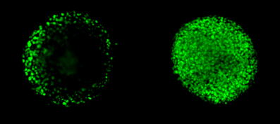 Image: NCI-H2170 spheroids approximately 250 µm in diameter labeled with nuclear stain. Left is in PBS and right is the same spheroid after clearing with Visikol HISTO-M. Image courtesy of Visikol.
Included in Corning Life Sciences’ comprehensive portfolio of 3D cell culture products are extracellular matrices for encapsulating cells within an ideal 3D growth environment, specialized microplates that allow cells to self-assemble into suspension colonies, and a range of permeable supports that mimic 3D microenvironments for a diverse set of cell culture applications. “Carefully optimized for generating, culturing, and assaying uniformly sized 3D multicellular spheroids, our spheroid microplates have an innovative U-shaped well geometry, clear bottom, and opaque side walls that eliminate the need for transfer steps,” explains Lynsey Willetts, cell culture business director at Corning Life Sciences. “Spheroid transfer can introduce variability to an assay, but by using our spheroid microplates in conjunction with a clearing agent such as Visikol and a pin-tool or liquid-handling equipment for solution changes to avoid disturbing the cultures, researchers can achieve clearer imaging and greater experiment reproducibility.”
Novel tools
3D cellular co-cultures are often used to study the interplay between different cell types, such as that which occurs during tumor metastasis or the recruitment of non-malignant cells to the tumor microenvironment. “Our Transwell permeable supports can easily be used to compartmentalize assays,” says Willetts, “and can be combined with extracellular matrices or spheroid microplates for greater flexibility. For example, an invasion assay could include 3D cancer cell cultures in a spheroid microplate with Transwell inserts, during which the proportion and behavior of cells migrating through the membrane to get to a higher concentration of attractant would be monitored.”
Designed for microfluidics-perfused growth of vessels, tubules, and organoids in 3D cell culture, the OrganoPlate® from Mimetas is a microplate format with tightly controlled fluid flow that is easily used for high-level imaging and can be incorporated into standard laboratory workflows without difficulty. “OrganoPlate is compatible with phase contrast, fluorescent and confocal imaging, and can also readily be used for time-lapse, since the bottom of the plate is made of high-quality, coverslip-thickness glass,” explains Bob Ronden, product manager. “Our 2-channel format features 96 independent culture cells, each supporting one in-gel culture and a perfusion channel, while a 3-channel version allows apical and basal access to epithelial and endothelial tubules.”
Also facilitating the creation of organized co-cultures without the requirement for artificial membranes, OrganoPlate has been used in a wide variety of applications, including the culture of perfused human renal proximal tubules, the growth of blood vessels, and the differentiation of iPS neurons. “We’re continually extending the capabilities of OrganoPlate,” adds Ronden. “For example, we recently developed a unique barrier integrity assay during which the tubule is perfused with a fluorescent dye. Using a standard fluorescent microscope, we were able to monitor dye movement through the barrier tissue and could quantify the barrier integrity by comparing the fluorescent intensity in the ECM channels to the intensity in the perfusion channels with endothelial or epithelial tubules. Every OrganoPlate user will have access to this protocol.”
Biocompare’s Cell Culture Search Tool
Find, compare and review cell culture
tools from different suppliers Search
MicroBrain® 3D Assay Ready plates from StemoniX are a human iPSC-derived cortical spheroid screening platform introduced to the 3D cell culture market in 2017 and already seeing considerable utility. Supplied in a pre-plated format, each well contains a single mature spheroid composed of neurons and astrocytes. “Through our adherence to strict manufacturing protocols we’re able to generate spheroids that demonstrate uniform size and morphology, exhibiting less than 3% variation in diameter,” says Ryan Gordon, vice president of business development and commercialization. “High-volume manufacturing addresses many common sources of variability, providing the pharmaceutical industry with a robust, reproducible, functional screening assay that is suitable to high-throughput formats.”
“The main application of microBrain® 3D Assay Ready plates is in the assessment of various drugs on the neural system,” adds Gordon. “The fields of neurotoxicology and neurological drug discovery are extremely active, and we’ve created a physiologically relevant human system to help pharmaceutical companies identify the safest and most efficacious drugs before human clinical testing.” Using a FLIPR Tetra® to image the calcium transport that accompanies neuronal firing, researchers can identify compounds that modulate the functional activity of the neural spheroids in StemoniX microBrain® 3D Assay Ready plates in their toxicity and efficacy research. High-content imaging can also be performed to detect neurological markers such as Synapsin I and Aquaporin 4.
The development of technologies such as StemoniX’ MicroBrain 3D Assay Ready plates has hugely increased the accessibility of 3D cell culture to the research community. Tried and trusted reagents like Visikol HISTO-M are also seeing additional use, although Johnson cautions that the most significant shortcoming with 3D cell culture today is the lack of consensus regarding the models themselves. “There are not yet standardized or generally accepted practices for generating and validating 3D cell culture models,” he says, “and instead this space is nascent. It is likely that in the future large companies will begin to democratize models to allow any researcher to easily generate their own model and validate it for a specific use case and application. In fact, we’re already starting to see this from companies such as Thermo Fisher Scientific with their new 3D cell culture resources and the NIH with their retina organoid competition.”
Ronden adds that for 3D imaging, the balance between speed, resolution, data storage, and sensitivity should always be considered. “While 3D cell culture allows you to collect a lot more information about your cultures, proper analysis of 3D datasets may take more time than their 2D counterparts. Handling these datasets should be well organized, especially when screening is of interest.”