Transfection is a core technique in cell biology research. Widespread laboratory applications such as recombinant protein expression and RNA interference owe their prevalence to advancements in chemical transfection methods.
Transfection experiments are often a tug-of-war between attaining efficient gene delivery and minimizing cellular toxicity. Transfection reagents are commonly comprised of cationic lipids and/or polymers that complex with negatively charged nucleic acids and promote cell entry via endocytosis. To avoid intracellular degradation, these transfection complexes must then escape from the endosome and deliver the nucleic acid to the cytoplasm (mRNA, siRNA) or the nucleus (plasmid DNA). Membrane-active properties of a reagent are desirable in that they enable the transfection complex to penetrate the plasma and endocytic membranes, but this often comes at the cost of eliciting a cytotoxic response. Few reagents on the market are able to simultaneously achieve high transfection efficiency while maintaining low toxicity.
Assessing Cellular Toxicity
The extent of cellular toxicity caused by transfection is influenced by the reagent and the nature of the cells. In particular cell types, dramatic toxicity can be directly visualized using microscopy (Figure 1, Reagent C). However, some cell types are more resilient, and although toxicity may not be immediately apparent, it can adversely affect experimental results. The cytotoxic effects of transfection are detrimental, as it can spur the nonspecific activation of certain genes that interfere with the experimental read-out. Certain commonly used liposomal transfection reagents are particularly known to activate stress response pathways that might affect cell cycle regulation and/or metabolic signaling. Cross talk between common signaling pathways may further confound the interpretation of results.
More sensitive detection of toxicity can be performed using assays for membrane leakiness (e.g., lactate dehydrogenase (LDH) release), viability (e.g., cellular staining for markers such as calcein-AM and propidium iodide) and cellular proliferation (e.g., MTT). The time point for each toxicity assay should be determined carefully, as some transfection reagents cause immediate cellular damage whereas others have more long-term effects. Detection at 24 hours post-transfection sufficiently illustrates the toxicity profile of most transfection reagents.
Researchers can take several steps to minimize transfection associated toxicity:
- Choose a reliable reagent.
- Ensure ideal concentration of reagent and nucleic acid.
- Select optimal cell culture conditions.
Transfection Reagent Selection
Cellular toxicity is largely dependent on the transfection reagent and cell type, making it particularly imperative to select a transfection reagent that balances high-efficiency nucleic acid delivery and low cellular toxicity. Although the exact composition of most commercial transfection reagents is unavailable; commonly used liposomal reagents tend to be most cytotoxic and should be avoided. Transfection reagents that are a combination of lipids and polyamines are frequently the most gentle to cells. Serum compatible transfection reagents are also preferred both for eliminating media changes post-transfection and maintaining cellular health.
Concentration of Reagent and Nucleic Acid
The concentration of transfection reagent and nucleic acid is critical to maximizing transfection efficiency while minimizing cellular toxicity. Cell types vary greatly in their sensitivity to the delivery and dosage of exogenous nucleic acid and reagent. Because of the complex and diverse formulations of commercially available transfection reagents, each reagent will need to be titrated on individual cell types. Testing at least three reagent levels is recommended. An excess of plasmid DNA can also contribute to cellular toxicity, and through empirical testing we have determined that a good starting point is one microgram per well of a 12-well plate. This results in maximum protein expression as well as minimal toxicity.
Cell Culture Considerations
Overall cellular health is another key parameter that influences transfection efficiency and toxicity. Highest transfection efficiencies are obtained when cells are actively dividing. This is particularly important for plasmid DNA transfections in which the nuclear envelope breakdown during cell division aids in uptake of the nucleic acid. For siRNA and mRNA transfections, the nucleic acid is functional in the cytoplasm; therefore, cell division is less critical. Additionally, the density of the cells at the time of transfection is essential to maintaining the health of the culture. The optimal confluency at the time of transfection is 60% to 80% for adherent cells. Transfection of cells at lower densities often leads to cellular toxicity because of an increase in the number of complexes delivered per cell.
Conclusion
Understanding the impact of transfection mediated toxicity is important for every researcher. The toxicity profile is largely dependent on the transfection reagent and cell type. Choosing a transfection reagent that balances high-efficiency nucleic acid delivery and cellular toxicity is imperative to achieve reliable experimental results.
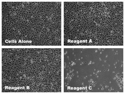
Figure 1. HeLa cells were imaged 24 hours post-transfection using three commercially available transfection reagents (A-C) at the recommended starting conditions for each product (3:1 reagent-to-DNA ratio).
Laura Juckem, Ph.D., is the R&D Group Leader at Mirus Bio.
Related Products from: Mirus Bio